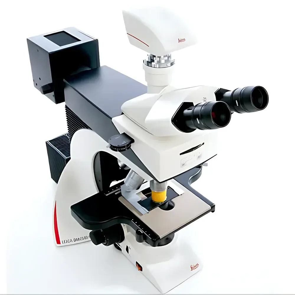
Leica DM2500 Biological Microscope
| Brand | Leica |
|---|---|
| Origin | Germany |
| Model | DM2500 |
| Illumination | 12 V / 100 W halogen transmitted-light source |
| Objective Turret | 6-position or 7-position manual turret |
| Eyepieces | 10× widefield, 22–25 mm field number |
| Observation Modes | Brightfield, Darkfield, Phase Contrast, Fluorescence, Polarization, Differential Interference Contrast (DIC) |
| Fluorescence Filter Changer | 5-position rotary filter cube holder |
| Tube Options | Binocular or trinocular head |
| Ergonomics | Height-adjustable focus knobs, tilting observation tube, modular stage controls |
Overview
The Leica DM2500 is a high-performance upright biological microscope engineered for demanding applications in pathology, clinical diagnostics, cell biology, hematology, and fundamental life science research. Built on Leica Microsystems’ decades-long legacy in precision optical design, the DM2500 employs an advanced Köhler illumination system centered around a stabilized 12 V / 100 W halogen lamp—delivering uniform, high-intensity transmitted light essential for quantitative brightfield analysis, unstained specimen visualization (e.g., live cells, blood smears), and high-contrast contrast-enhancement techniques including DIC and phase contrast. Its rigid, thermally stable mechanical platform ensures long-term optical alignment integrity, while its modular architecture supports seamless integration of specialized contrast methods without compromising parfocality or mechanical reproducibility. Designed for compliance with ISO 10993 biocompatibility standards for medical device components and aligned with GLP/GMP laboratory infrastructure requirements, the DM2500 serves as a validated core imaging platform in regulated diagnostic environments.
Key Features
- 7-position manual objective turret accommodating full-line Leica HC PL FLUOTAR, HCX PL FLUOTAR, and HC PL APO objectives—including plan achromat, semi-apochromat, and apochromat variants with correction for chromatic and spherical aberration across visible and near-UV spectra.
- DIC-ready optical path with integrated Nomarski prism sliders and strain-free polarizers; optimized for sub-micron topographic resolution in unstained, hydrated specimens without fixation or staining.
- 5-position rotary fluorescence filter cube changer enabling rapid switching between DAPI, FITC, TRITC, Cy5, and custom excitation/emission configurations—fully compatible with Leica’s zero-pixel-shift emission path design for spatially accurate multi-channel registration.
- Ergonomic height-adjustable coarse/fine focus knobs with dual-speed gearing (1 mm per revolution coarse, 10 µm per revolution fine), supporting extended-duration microscopy sessions while minimizing operator fatigue.
- Modular trinocular head with 20° inclined viewing angle, interchangeable widefield 10× eyepieces (22–25 mm field number), and C-mount port for standardized CCD, sCMOS, or DSLR camera coupling.
- Adjustable mechanical stage with low-profile X-Y translation, optional vernier scale, and compatibility with motorized stage controllers for semi-automated slide scanning workflows.
Sample Compatibility & Compliance
The DM2500 accommodates standard 1–2 mm thick glass slides, Petri dishes (up to 35 mm diameter), and culture flasks via optional stage adapters. Its open optical architecture permits use with immersion media (water, glycerol, oil) across 4×–100× magnifications. All optical components comply with ISO 8578 (microscope performance testing) and DIN EN 61000-6-3 (EMC emissions). The instrument meets IEC 61010-1 safety requirements for laboratory equipment and supports audit-ready documentation packages for FDA 21 CFR Part 11–compliant digital imaging systems when paired with Leica LAS X software and certified storage protocols.
Software & Data Management
The DM2500 interfaces natively with Leica Application Suite (LAS) X v3.7+ via USB 3.0 or GigE Vision. LAS X provides calibrated measurement tools (area, length, intensity profiling), multi-channel fluorescence overlay, Z-stack acquisition, time-lapse synchronization, and export to TIFF, OME-TIFF, or HDF5 formats. Metadata embedding includes objective ID, exposure settings, filter position, and user-defined annotations—enabling traceability in ISO/IEC 17025-accredited laboratories. Integration with third-party platforms (e.g., HALO®, Visiopharm®, QuPath) is supported through standardized OpenMicroscopy Environment (OME) APIs.
Applications
- Diagnostic histopathology: Routine H&E, special stains, and immunohistochemistry evaluation under brightfield and polarization modes.
- Live-cell imaging: Phase contrast and DIC observation of primary cultures, organoids, and suspension cells without phototoxicity from intense excitation.
- Hematology: Differential white blood cell counts, platelet morphology assessment, and malaria parasite detection in Giemsa-stained peripheral smears.
- Fluorescence cytogenetics: Rapid karyotyping using chromosome-specific FISH probes with minimal crosstalk due to high-transmission dichroic filters.
- Materials-assisted biology: Birefringence analysis of collagen fibers, starch granules, or crystalline drug formulations under polarization optics.
FAQ
Is the Leica DM2500 compliant with regulatory standards for clinical diagnostics?
Yes—the platform conforms to ISO 13485 design control principles and supports validation documentation packages required for CE-IVD and FDA 510(k) submission pathways when deployed with approved accessories and software modules.
Can the DM2500 be upgraded for automated scanning or whole-slide imaging?
While the base model is manually operated, it accepts Leica’s DM2500-Motorized Stage Kit and is compatible with third-party slide scanners via standardized stage coordinate mapping and TTL trigger interfaces.
What is the maximum numerical aperture supported in fluorescence mode?
When equipped with Leica HC PL APO 100×/1.40 Oil objectives and appropriate filter sets, the system achieves effective NA ≥ 1.35 in epifluorescence configuration, ensuring optimal signal-to-noise ratio for dim fluorophores.
Does the 100 W halogen illumination generate excessive heat during prolonged DIC observation?
The integrated IR-cut filter and air-cooled lamp housing limit thermal drift to <0.5 µm/min at the focal plane over 60-minute sessions—validated per ISO 9343 thermal stability test protocols.


