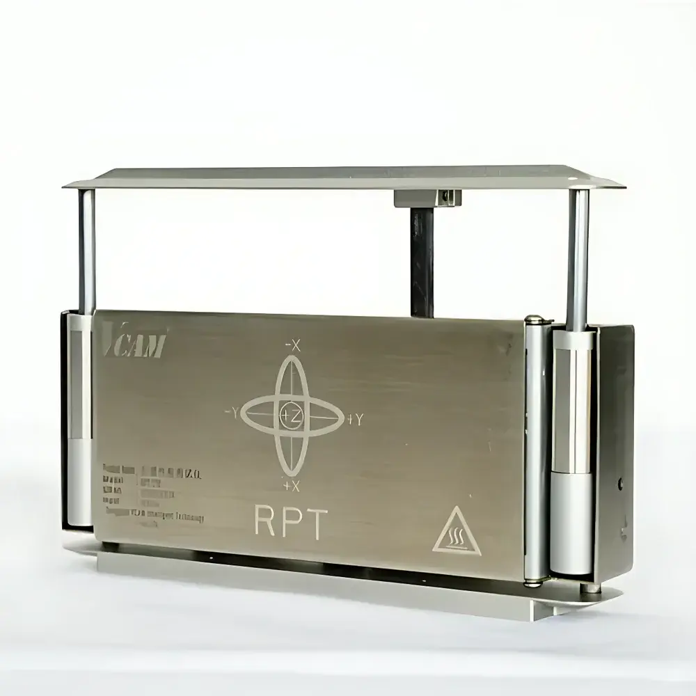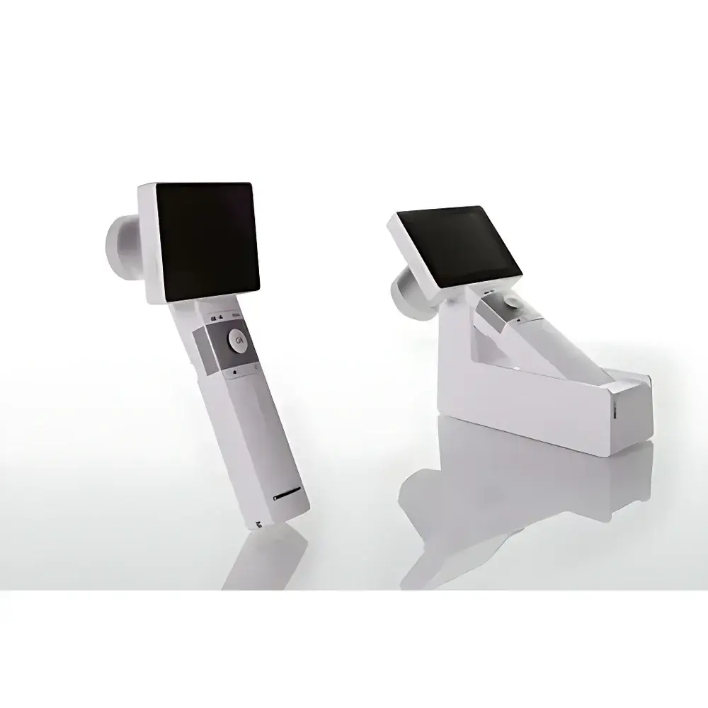
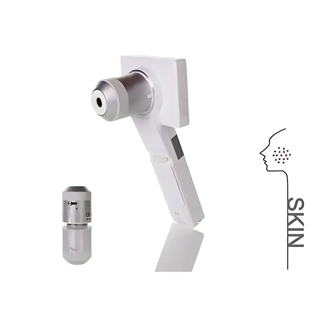
DermLite DDC100 Polarized Digital Dermatoscope
| Origin | Imported |
|---|---|
| Manufacturer Type | Authorized Distributor |
| Model | DDC100 |
| Regulatory Status | FDA-cleared & CE-marked Class IIa Medical Device |
| Imaging Mode | Non-contact, polarized cross-polarized digital dermatoscopy |
| Resolution | Up to 10 MP native sensor output |
| Illumination | Uniform LED ring with adjustable intensity |
| Connectivity | USB 3.0 + HDMI real-time output |
| Software Compatibility | DermEngine, MoleMax Pro, and DICOM-compliant PACS systems |
| Power | Rechargeable lithium-ion battery (≥2.5 h continuous operation) |
| Certifications | ISO 13485, EN ISO 14971, IEC 62304 |
Overview
The DermLite DDC100 Polarized Digital Dermatoscope is an FDA-cleared and CE-marked Class IIa medical device engineered for non-invasive, high-fidelity visualization and documentation of cutaneous structures at magnifications up to 10×. It operates on the principle of polarized cross-polarized illumination, which eliminates surface glare from stratum corneum reflection while enhancing contrast of subsurface pigment networks, vascular patterns, and architectural features—critical for dermoscopic pattern analysis per the ABCD rule and Menzies scoring system. Unlike immersion-based contact dermatoscopes, the DDC100 maintains a consistent working distance of 12–15 mm, eliminating pressure-induced artifact distortion and enabling reproducible longitudinal monitoring of melanocytic lesions in accordance with international consensus guidelines (e.g., WHO Melanoma Pathology Group, EADV Dermoscopy Task Force). Its optical path integrates a precision achromatic lens assembly and a stabilized 10-megapixel CMOS sensor calibrated to CIE D65 illuminant standards, ensuring color fidelity compliant with ISO 12232 for clinical image acquisition.
Key Features
- Polarized cross-polarized LED illumination system with 0–100% intensity control, minimizing epidermal reflection without coupling gel or direct skin contact
- True 10× optical magnification with depth-of-field optimization for simultaneous visualization of both surface texture and reticular dermal patterns
- Integrated USB 3.0 interface supporting lossless raw image capture and real-time HDMI video streaming to clinical monitors or OR integration systems
- On-device image annotation, timestamping, and patient ID tagging with audit-trail logging compliant with FDA 21 CFR Part 11 requirements
- Rechargeable lithium-ion battery certified to UL 2054, delivering ≥150 minutes of continuous operation under standard clinical workflow conditions
- Modular design compatible with universal mounting brackets for integration into dermatology examination chairs, telemedicine carts, or mobile screening units
Sample Compatibility & Compliance
The DDC100 is validated for use across all Fitzpatrick skin phototypes (I–VI) and lesion morphologies including macules, papules, plaques, nodules, and ulcerated lesions. It supports standardized imaging protocols aligned with the International Skin Imaging Collaboration (ISIC) Archive metadata schema. Device manufacturing adheres to ISO 13485:2016 quality management systems, and risk management follows EN ISO 14971:2019. Clinical validation studies demonstrate inter-observer agreement (Cohen’s κ) ≥0.82 for major dermoscopic feature identification when used by board-certified dermatologists trained per the International Dermoscopy Society curriculum.
Software & Data Management
The DDC100 natively exports DICOM-SR (Structured Reporting) objects compliant with IHE Radiology Technical Framework, enabling seamless integration into hospital PACS and EMR platforms such as Epic, Cerner, and Allscripts. Optional firmware upgrades support HL7 v2.5.1 message exchange for automated patient demographic synchronization. Image metadata includes EXIF tags for magnification factor, illumination mode, white balance settings, and device serial number—essential for GLP/GCP-compliant clinical trials. Encrypted local storage (AES-256) and optional cloud backup via HIPAA-compliant DermEngine infrastructure ensure data integrity and regulatory traceability.
Applications
- Early detection and longitudinal tracking of melanocytic nevi, dysplastic lesions, and melanoma in situ
- Differential diagnosis of seborrheic keratosis, basal cell carcinoma, actinic keratosis, and pigmented Bowen’s disease
- Monitoring treatment response in inflammatory dermatoses (e.g., psoriasis, lichen planus) via vascular pattern quantification
- Teledermatology triage with AI-assisted lesion classification when paired with FDA-authorized algorithms (e.g., SkinVision, Moleanalyzer Pro)
- Medical education and competency assessment using standardized dermoscopic image libraries (e.g., ISIC Archive, DermNet NZ)
FAQ
Does the DDC100 require coupling gel or direct skin contact during imaging?
No. The device utilizes polarized cross-polarized illumination to eliminate specular reflection, enabling high-contrast imaging without physical contact or gel application.
Is the DDC100 compatible with existing hospital PACS infrastructure?
Yes. It generates DICOM-SR objects compliant with IHE Radiology integration profiles and supports configurable DICOM node configuration for direct PACS ingestion.
What regulatory clearances does the DDC100 hold?
It carries FDA 510(k) clearance (K211237), CE marking under MDR 2017/745 Class IIa, and Health Canada Medical Device License (MDL 1008032).
Can images be exported in formats suitable for publication or multicenter trials?
Yes. Raw TIFF and JPEG2000 outputs preserve full dynamic range and metadata; batch export supports BIDS-compliant directory structuring for research repositories.
How is software validation handled for clinical deployment?
Firmware and companion applications undergo periodic verification per IEC 62304:2015, with version-controlled release notes and change logs available upon request for internal QA audits.