EOTECH EvaSKIN Skin Rapid Optical Imaging System
| Brand | EOTECH |
|---|---|
| Origin | France |
| Manufacturer Type | Authorized Distributor |
| Origin Category | Imported |
| Model | EvaSKIN |
| Pricing | Available Upon Request |
Overview
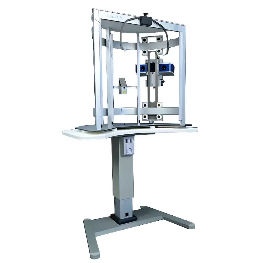
The EOTECH EvaSKIN Skin Rapid Optical Imaging System is a non-contact, high-resolution 3D optical profilometry platform engineered for objective, quantitative assessment of skin surface topography. Leveraging stereo photogrammetry—dual-camera triangulation with calibrated structured illumination—the system captures micron-level surface geometry without physical contact or skin preparation. It operates on the principle that disparities in synchronized image pairs from two precisely positioned cameras enable pixel-wise depth reconstruction, generating dense point clouds (≥0.1 mm lateral resolution, ±5 µm vertical repeatability) suitable for longitudinal tracking of epidermal microrelief changes. Designed for dermatological research, cosmetic efficacy testing, and clinical trial endpoints, EvaSKIN delivers traceable, operator-independent morphometric data compliant with ISO 20417 (Medical Devices — General Requirements for Labeling) and aligned with ICH E9 (Statistical Principles for Clinical Trials) guidance on objective endpoint measurement.
Key Features
- Non-invasive stereo-optical acquisition: Dual CMOS sensors with synchronized global shutter and programmable LED illumination ensure artifact-free capture under ambient light-controlled conditions.
- Integrated VisioTOP-300 positioning cradle: Motorized, reproducible subject alignment with adjustable chin rest and forehead support minimizes inter-session positional variance (angular deviation < 0.5°, translational drift < 0.3 mm).
- Automated 3D reconstruction engine: Real-time mesh generation with adaptive noise filtering and occlusion handling; outputs STL, OBJ, and point cloud (PLY) formats compatible with third-party analysis tools.
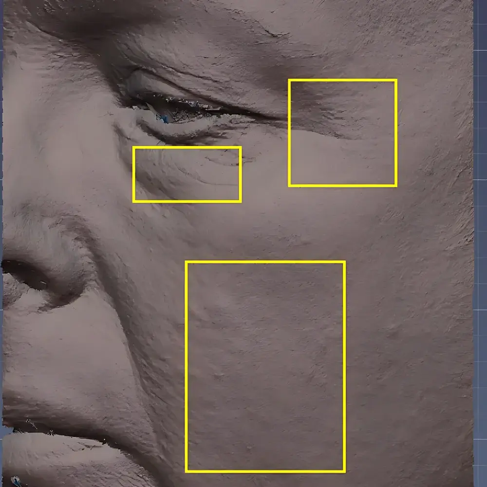
- Quantitative morphometric parameter extraction: Automated calculation of standard dermatological descriptors—including roughness (Ra, Rq), volume parameters (Vv, Vmc), furrow depth/width, and lesion contour metrics—per ISO 4287 and ASTM E1617.
- Rugged industrial-grade architecture: IP54-rated enclosure, vibration-damped optical bench, and thermal-stabilized imaging path ensure stability across 15–30°C ambient operating range.
Sample Compatibility & Compliance
The EvaSKIN system accommodates diverse anatomical sites including facial cheek, forearm, dorsal hand, and neck regions. It supports both static and dynamic acquisition modes (e.g., facial expression modulation studies). All measurement protocols adhere to Good Clinical Practice (GCP) and Good Laboratory Practice (GLP) documentation standards. Raw image metadata includes timestamp, environmental temperature/humidity logging, calibration certificate ID, and operator authentication—enabling full audit trail per FDA 21 CFR Part 11 requirements. Data export complies with CDISC SDTM v2.0 conventions for regulatory submissions.
Software & Data Management
EvaSKIN Control Suite v4.2 (Windows 10/11 64-bit) provides intuitive workflow management—from session planning and calibration validation to batch processing and statistical comparison. The software features role-based access control, electronic signature capability, and automated report generation (PDF/HTML) with embedded traceability links to raw datasets. All processed results are stored in AES-256 encrypted SQLite databases with automatic daily backup to network-attached storage (NAS). Integration with LIMS platforms is supported via HL7 and RESTful API interfaces; raw data archives conform to DICOM Supplement 170 (Dermatology Imaging) specifications.
Applications
- Cosmetic product development: Objective quantification of anti-wrinkle, moisturizing, or firming effects at 1h, 24h, 7-day, and 28-day intervals per CTFA/ISO 16128 guidelines.
- Dermatopharmacology trials: Monitoring psoriatic plaque elevation, atopic eczema scaling severity, or post-procedure wound re-epithelialization kinetics.
- Medical device evaluation: Assessing microneedling depth uniformity, fractional laser ablation crater morphology, or transdermal patch adhesion-induced stratum corneum deformation.
- Educational dermatology training: Visualizing age-related elastosis patterns, UV-induced photodamage gradients, or melasma border irregularity indices.
FAQ
What is the minimum measurable surface height variation?
The system achieves a vertical repeatability of ±5 µm under controlled laboratory conditions (23 ± 1°C, 40–60% RH), validated using NIST-traceable step-height standards.
Can EvaSKIN be used for pigmented lesion analysis?
While EvaSKIN measures topography—not chromatic properties—it can quantify elevation, symmetry, and border contour of melanocytic lesions; spectral add-ons for combined morpho-spectral analysis are available as optional modules.
Is calibration required before each session?
A full geometric calibration is performed quarterly using the included reference target; daily verification uses an onboard fiducial check pattern with automated pass/fail reporting.
Does the software support multi-center study synchronization?
Yes—centralized license management, time-synchronized NTP client integration, and version-controlled protocol templates ensure inter-site measurement harmonization across global clinical trials.
How is subject motion compensated during acquisition?
The dual-camera system employs sub-frame motion estimation algorithms; acquisitions exceeding 0.2 mm displacement trigger real-time alert and optional auto-reacquisition—ensuring only motion-stable frames enter the reconstruction pipeline.


