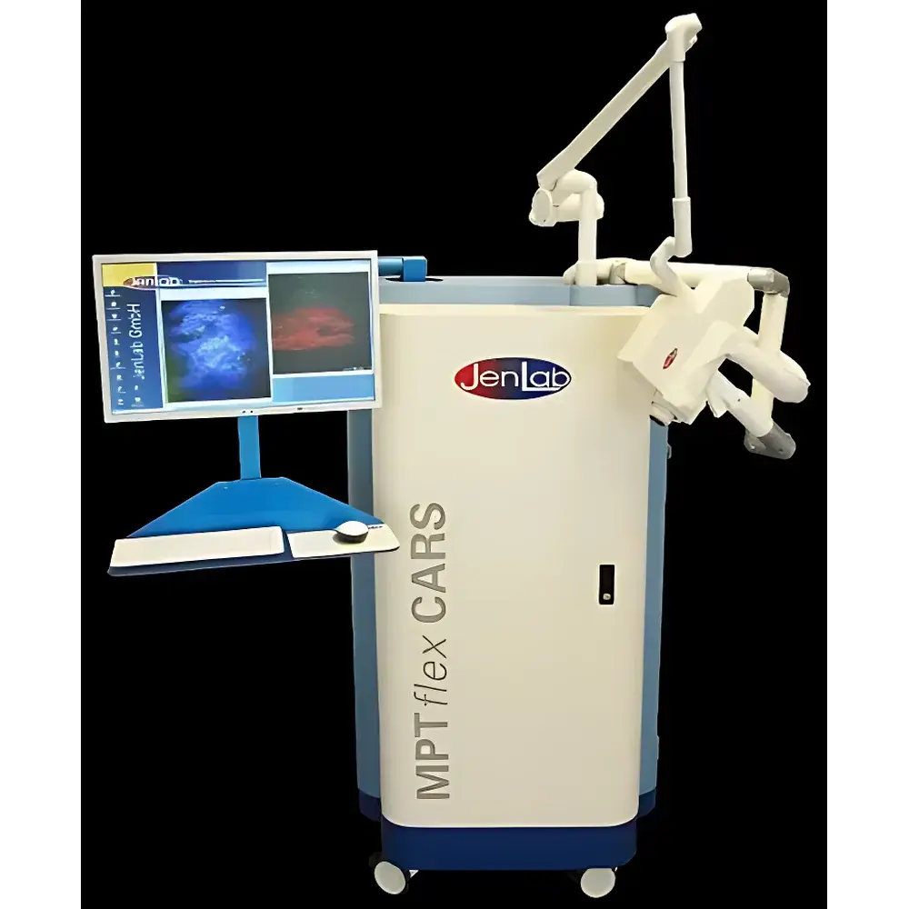
CK MPT flex Multiphoton Tomography System for Skin Imaging
| Brand | CK |
|---|---|
| Origin | Germany |
| Manufacturer Status | Authorized Distributor |
| Origin Category | Imported |
| Model | MPT flex |
| Pricing | Upon Request |
Overview
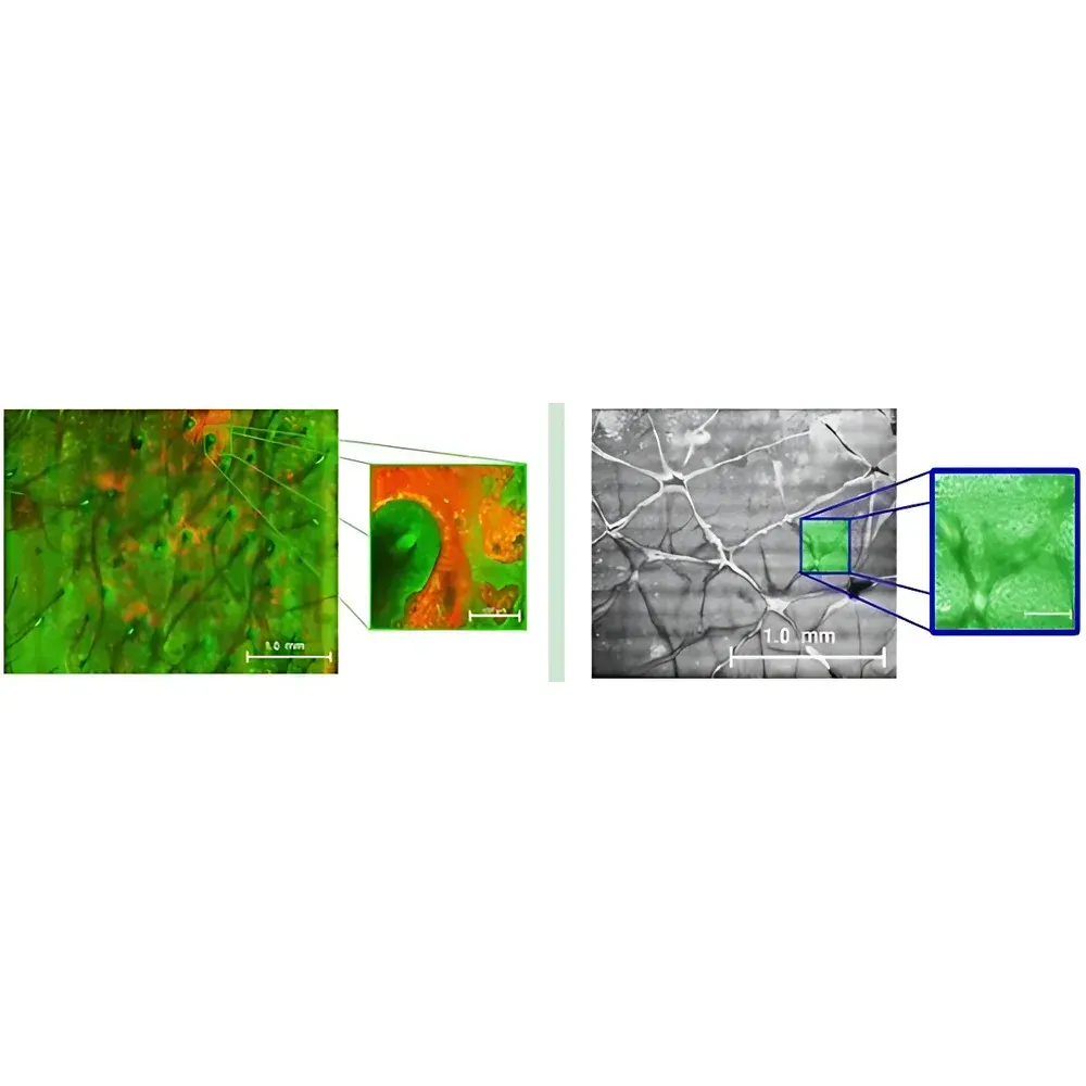
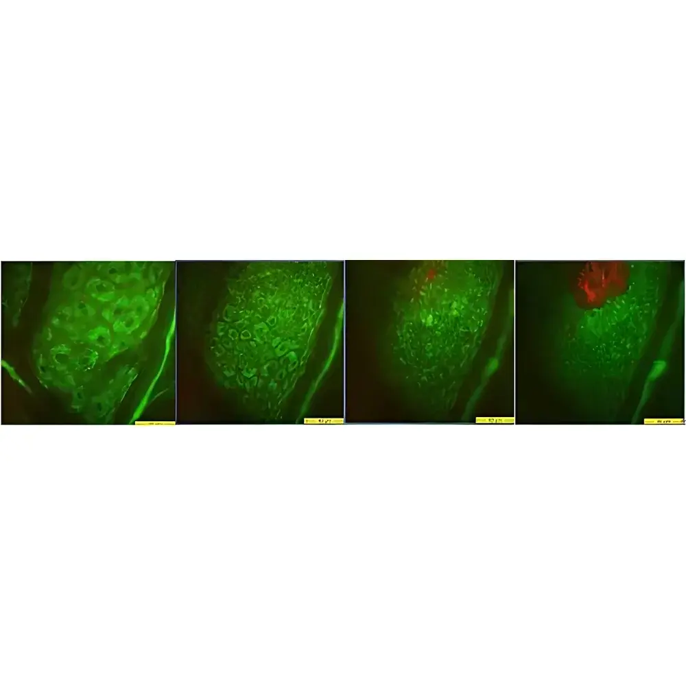
The CK MPT flex Multiphoton Tomography System is a high-resolution, non-invasive optical imaging platform engineered for label-free, in vivo 3D visualization of human and murine skin microstructure at subcellular resolution. Based on femtosecond near-infrared (NIR) laser excitation (typically 760–800 nm), the system leverages multiphoton excitation fluorescence (MPEF) and second-harmonic generation (SHG) to generate intrinsic contrast from endogenous fluorophores—including NAD(P)H, FAD, melanin, keratin, and collagen—without requiring exogenous dyes or tissue sectioning. This principle enables real-time, depth-resolved assessment of epidermal morphology, dermal extracellular matrix architecture, melanocyte distribution, and vascular networks down to ~250 µm depth with lateral resolution <0.5 µm and axial resolution <1.5 µm. Designed specifically for dermatological translational research, the MPT flex complies with IEC 60601-1 safety standards for medical electrical equipment and supports clinical-grade imaging protocols aligned with ISO 13485–certified quality management systems.
Key Features
- Femtosecond NIR laser source (Ti:Sapphire oscillator, pulse width <100 fs, repetition rate ~80 MHz) optimized for deep-tissue penetration and minimal photodamage
- Integrated galvanometric scanning unit enabling high-speed raster scanning (up to 12 frames/s at 512 × 512 pixels) with precise z-stack acquisition
- Dual-channel detection path with spectral separation for simultaneous MPEF and SHG signal collection using high-quantum-efficiency GaAsP photomultiplier tubes
- Motorized XYZ stage with micrometer-level precision positioning and ergonomic patient interface for reproducible longitudinal imaging sessions
- Modular design supporting interchangeable objectives (20×/0.75 NA, 40×/0.95 NA water-immersion) and optional add-ons including time-resolved fluorescence lifetime (FLIM) module and polarization-resolved SHG
- Compliant with laser safety Class IV requirements and equipped with interlocked enclosure, beam shutter, and real-time power monitoring per EN 60825-1
Sample Compatibility & Compliance
The MPT flex accommodates intact human skin (volunteer and biopsy specimens), ex vivo porcine and murine tissue models, 3D skin equivalents (e.g., reconstructed epidermis, full-thickness organotypic cultures), and live anesthetized small-animal preparations. It supports imaging under physiological hydration conditions using coupling gel or immersion medium. All hardware and firmware meet CE marking requirements for in vitro diagnostic (IVDR) devices where applicable. Data acquisition workflows are compatible with Good Laboratory Practice (GLP) documentation standards; audit trails, user access control, and electronic signature support can be implemented via optional software modules compliant with FDA 21 CFR Part 11.
Software & Data Management
Acquisition and analysis are performed using proprietary MPT Image Suite v5.x, a Windows-based application providing real-time image preview, automated focus tracking, multi-parameter intensity quantification (e.g., epidermal thickness, collagen fiber orientation index, melanin density maps), and batch processing for longitudinal datasets. Raw data are stored in vendor-neutral TIFF or HDF5 format with embedded metadata (laser power, scan speed, objective ID, timestamp). Export options include 3D surface rendering (STL), volumetric projections (MP4, AVI), and tabular CSV outputs for statistical analysis in MATLAB, Python (via open-source MPT-Reader libraries), or commercial platforms such as Imaris or Fiji/ImageJ. Software updates follow a documented change control process aligned with ISO 14971 risk management principles.
Applications
- Early detection and morphometric characterization of melanocytic lesions, including dysplastic nevi and stage I melanoma, through quantitative assessment of melanosome distribution and keratinocyte atypia
- Evaluation of chronic inflammatory dermatoses (e.g., psoriasis, atopic dermatitis) by mapping T-cell infiltration patterns and epidermal barrier integrity
- Preclinical assessment of topical drug delivery kinetics and transdermal penetration pathways using intrinsic fluorescence contrast
- Longitudinal monitoring of photoaging and chronological aging biomarkers—including elastosis, collagen fragmentation, and mitochondrial redox state (NADH/FAD ratio)
- In situ validation of 3D skin tissue engineering constructs, focusing on stratification, basement membrane formation, and functional melanocyte integration
- Stem cell engraftment tracking in murine wound-healing models via label-free autofluorescence signatures and niche interaction analysis
FAQ
Is the MPT flex suitable for regulatory submission studies (e.g., FDA IND or EMA CTA)?
Yes—when deployed within a validated environment with documented SOPs, instrument qualification (IQ/OQ/PQ), and electronic record controls, the system supports data generation acceptable for regulatory filings.
Can the system be integrated into existing microscopy core facilities?
Yes—the MPT flex features standard Ethernet and USB 3.0 interfaces, supports DICOM export via optional gateway, and operates within typical core lab environmental specifications (20–25°C, <60% RH, vibration-isolated benchtop).
What maintenance is required beyond routine calibration?
Annual laser power verification and PMT gain recalibration are recommended; full optical alignment and dispersion compensation checks are performed biannually by CK-certified field service engineers.