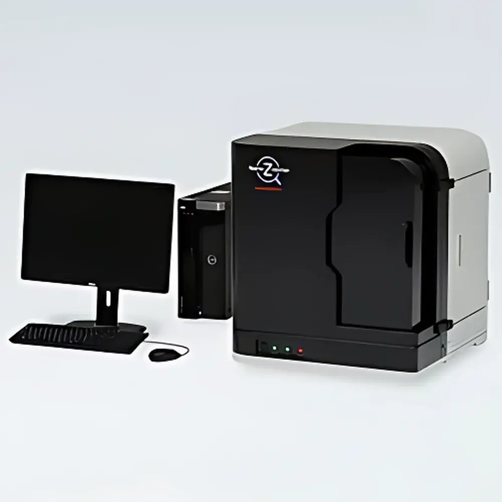
Hamamatsu NanoZoomer S60 Digital Whole Slide Scanner C13210-01
| Brand | Hamamatsu |
|---|---|
| Origin | Japan |
| Manufacturer | Hamamatsu Photonics K.K. |
| Product Type | Research-Use-Only (RUO) Digital Pathology Scanner |
| Model | C13210-01 |
| Imaging Modes | Brightfield and Fluorescence |
| Scan Field | Full-slide panoramic imaging |
| Throughput | High-throughput (up to 60 slides per load) |
| Operation | Fully automated |
| Form Factor | Benchtop |
| Optical Resolution | 0.46 µm/pixel (20×), 0.23 µm/pixel (40×) |
| Slide Capacity | 60 standard slides (26 mm × 76 mm, 0.9–1.2 mm thickness) or 30 dual-format slides (52 mm × 76 mm, optional) |
| Scan Speed | ~60 s per 15 mm × 15 mm field at 20× |
| Objective | 20× (N.A. 0.75), user-selectable 20×/40× scanning mode |
| Focus Method | Pre-focus map + Z-stack acquisition capability |
| Barcode Reader | Integrated 1D barcode scanner (2D optional) |
| Output Format | JPEG-compressed pyramidal TIFF-compatible tiles with embedded slide metadata |
| Power Supply | AC 100–240 V, 50/60 Hz |
| Power Consumption | ~225 VA |
| Regulatory Status | RUO in USA, Canada, Israel, Turkey |
Overview
The Hamamatsu NanoZoomer S60 (Model C13210-01) is a high-performance, benchtop digital whole slide scanner engineered for research-grade pathology imaging. Utilizing precision motorized stage control, high-numerical-aperture optics, and synchronized fluorescence excitation/emission filtering, the system captures spatially resolved, quantitative brightfield and fluorescence histopathological data across entire glass slides. Designed for reproducible, large-scale tissue digitization, it employs a tile-based scanning architecture with dynamic autofocus mapping and optional Z-stack acquisition—enabling robust optical sectioning of thick or unevenly mounted specimens. Unlike clinical IVD variants (e.g., S60v2MD), the C13210-01 is designated strictly for research use, complying with ISO 13485-aligned manufacturing processes while excluding FDA 510(k) or IVDR conformity for diagnostic interpretation.
Key Features
- Fully automated, unattended operation with 60-slide capacity using standardized cassette-based loading (20 slides × 3 cassettes) or dual-format support (30 slides × 3 cassettes for 52 mm × 76 mm slides)
- Dual-modality imaging: simultaneous brightfield and fluorescence scanning with hardware-synchronized filter wheels and LED-based excitation sources optimized for common fluorophores (DAPI, FITC, TRITC, Cy5)
- High-fidelity optical path: 20× objective (N.A. 0.75) delivering 0.46 µm/pixel resolution at 20× and 0.23 µm/pixel at 40× equivalent magnification—validated per ISO 19003:2021 for digital pathology image fidelity
- Efficient scan kinetics: ~60 seconds per 15 mm × 15 mm field at 20×; ~150 seconds at 40×—achieving sub-2-minute full-slide acquisition for standard 26 mm × 76 mm sections
- Integrated 1D barcode reader (2D optional) for automatic slide identification and metadata association; supports GS1-compliant barcoding standards
- Pre-focus map generation prior to scanning ensures consistent focus across heterogeneous tissue topographies; Z-stack functionality enables depth-resolved analysis of multi-layered samples
Sample Compatibility & Compliance
The NanoZoomer S60 accommodates standard microscope slides (26 mm × 76 mm, 0.9–1.2 mm thickness) and optionally accepts extended-format slides (52 mm × 76 mm). It is compatible with conventional H&E, immunohistochemical (IHC), and multiplex fluorescence-stained preparations—including those mounted with aqueous or permanent media. No proprietary slide coatings or adhesives are required. From a regulatory standpoint, the C13210-01 is classified as a Research Use Only (RUO) instrument under U.S. FDA guidance (21 CFR §809.3(f)) and Health Canada’s Medical Devices Regulations (SOR/98-282). It is not intended for clinical diagnosis, patient management, or therapeutic decision-making. In China, this model is registered as an in vitro diagnostic medical device (NMPA Registration No. pending public listing for C13210-01); however, its RUO labeling remains enforced outside certified IVD jurisdictions. The system adheres to IEC 61000-6-3 (EMC) and IEC 61000-6-2 (immunity) standards, and its mechanical design conforms to ISO 14155:2020 principles for laboratory equipment safety.
Software & Data Management
Controlled via Hamamatsu’s NDP.scan software (v3.x or later), the NanoZoomer S60 generates pyramidal, multi-resolution TIFF files compliant with OpenSlide and ASV-compatible viewers. Each output includes embedded EXIF metadata (objective, magnification, exposure time, illumination intensity, timestamp, slide ID) and supports DICOM-SR structured reporting when integrated with third-party PACS via HL7/FHIR middleware. Audit trails—including operator login, scan parameters, calibration logs, and error events—are retained locally and exportable in CSV format. While native software does not implement FDA 21 CFR Part 11 electronic signature controls, the system architecture permits integration with validated LIMS or ELN platforms that enforce role-based access, electronic signatures, and revision history tracking—supporting GLP and GCP-aligned workflows in academic and pharmaceutical research environments.
Applications
- Quantitative morphometric analysis of tumor microenvironments in preclinical oncology studies
- Longitudinal assessment of tissue biomarker expression across serial sections in translational neuroscience research
- High-content screening of organoid or 3D culture models using multiplex fluorescence protocols
- Training dataset generation for AI-based segmentation and classification algorithms (e.g., tumor grading, stromal infiltration scoring)
- Archival digitization of legacy histology collections with preservation of spectral fidelity for retrospective reanalysis
- Multi-institutional collaborative studies requiring standardized, vendor-neutral image exchange via OpenSlide or DICOM WSI profiles
FAQ
Is the NanoZoomer S60 C13210-01 approved for clinical diagnostics in the United States?
No. This model is designated Research Use Only (RUO) and is not cleared or approved by the U.S. FDA for diagnostic purposes.
Does the system support whole-slide fluorescence imaging with spectral unmixing?
It supports multi-channel fluorescence acquisition with discrete filter sets but does not include real-time spectral unmixing or hyperspectral detection capabilities.
Can the NanoZoomer S60 interface with existing hospital PACS systems?
Yes—via DICOM WSI conformance (supplement 145) when configured with optional DICOM gateway modules and validated network infrastructure.
What is the maximum tissue thickness supported for Z-stack acquisition?
The system supports Z-stack acquisition up to 50 µm total depth with user-defined step intervals (minimum 0.2 µm), subject to objective working distance and coverslip thickness constraints.
Is remote monitoring or cloud-based image upload natively supported?
NDP.scan provides local network sharing and scheduled export functions; cloud integration requires third-party middleware compliant with HIPAA, GDPR, or equivalent data sovereignty frameworks.
Related Products


