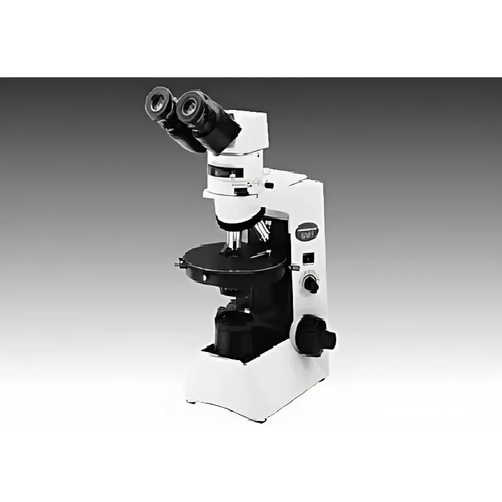
Olympus CX31-P Polarizing Microscope
| Brand | Olympus |
|---|---|
| Origin | Japan |
| Manufacturer | Olympus Corporation |
| Type | Imported Instrument |
| Model | CX31-P |
| Pricing | Upon Request |
Overview
The Olympus CX31-P is a compact, high-performance polarizing microscope engineered for rigorous qualitative and semi-quantitative birefringence analysis in materials science, geology, pharmaceuticals, and polymer research. Built upon the UIS2 (Universal Infinity System) optical platform, it delivers infinite-corrected optics with optimized chromatic and spherical aberration correction—essential for accurate interference figure observation and retardation measurement. The instrument supports standardized transmitted-light polarized light microscopy (PLM), including both plane-polarized and crossed-nicols configurations, as well as conoscopy via interchangeable Bertrand lens and swing-in condenser optics. Its rigid mechanical architecture minimizes stage drift and focus shift during extended observation or sequential imaging, ensuring measurement repeatability required for GLP-compliant documentation and ASTM E112/E45-compliant grain/phase characterization.
Key Features
- UIS2 infinity-corrected optical system with dedicated polarizing objectives: PLN4×P, ACHN-P, and UPLFLN-P series—designed to minimize strain-induced birefringence and maximize extinction contrast.
- Rigid monolithic frame with precision-machined focusing mechanism (25 mm coarse travel, 0.2 mm fine adjustment per revolution) and integrated rotating mechanical stage (360° rotation with engraved scale, ±0.1° vernier readout).
- Dual-purpose observation tube: binocular or trinocular configuration with fixed crosshair orientation—maintains alignment of reticle axes relative to vibration direction regardless of interpupillary adjustment.
- High-contrast polarizing accessories: redesigned polarizer/analyzer set with 180° rotation (2° incremental scale, 0.1° vernier resolution), enhanced numerical aperture condenser optimized for polarized illumination (EF value improved vs. prior generations).
- Modular imaging readiness: trinocular port compatible with C-mount digital cameras; supports time-stamped image capture, metadata embedding (magnification, objective ID, analyzer angle), and integration into regulated lab environments compliant with FDA 21 CFR Part 11 audit trail requirements.
Sample Compatibility & Compliance
The CX31-P accommodates standard 24 × 50 mm and 48 × 60 mm petrographic slides, as well as custom-thickness specimens up to 30 mm (with optional long-working-distance objectives). It meets ISO 8578:2017 (microscopes — requirements and tests) for mechanical stability and optical performance verification. All polarizing components are calibrated per ASTM D4124 (standard test method for birefringence of thermoplastic polymers) and support quantitative retardation assessment using λ-plate and quartz wedge compensators (optional). The system is suitable for USP microscopy validation protocols and routine QC in GMP-regulated pharmaceutical crystallinity testing.
Software & Data Management
While the CX31-P operates as a standalone optical instrument, its trinocular port enables seamless integration with Olympus cellSens™ Digital Imaging Software or third-party DICOM-compliant platforms. Captured images retain EXIF-style metadata—including analyzer angle, objective magnification, illumination intensity, and timestamp—enabling traceable correlation between optical conditions and analytical outcomes. Audit-ready log files comply with ALCOA+ principles (Attributable, Legible, Contemporaneous, Original, Accurate) and support electronic record retention per Annex 11 and 21 CFR Part 11 when deployed with validated software workflows.
Applications
- Mineral identification and optical mineralogy (interference color charting, optic sign determination, extinction angle measurement)
- Pharmaceutical solid-state characterization (polymorph screening, crystal habit analysis, stress-induced birefringence in tablet coatings)
- Polymer morphology assessment (spherulite size distribution, lamellar orientation, phase separation in blends)
- Forensic fiber analysis (birefringence magnitude and sign differentiation between synthetic and natural fibers)
- Quality control of liquid crystal displays (LCD) substrates and optical films
FAQ
Does the CX31-P support both orthoscopic and conoscopic observation?
Yes—the instrument includes a swing-in Bertrand lens and selectable condenser optics enabling both standard plane-polarized imaging and conoscopy for uniaxial/biaxial crystal analysis.
Is the analyzer angle digitally encoded or manually read?
The analyzer rotates mechanically with a vernier scale providing 0.1° resolution; digital encoding requires external motorized rotation stages (not included but compatible via third-party adapters).
Can the CX31-P be used for quantitative retardation measurements?
Yes—when paired with certified λ-plates (e.g., 530 nm first-order red plate) or quartz wedge compensators (sold separately), it supports semi-quantitative retardation estimation per ISO 10110-5 guidelines.
What is the maximum specimen thickness supported without modification?
Standard configuration accommodates specimens up to 1.2 mm thick under 40×P objective; thicker samples require long-working-distance objectives (e.g., LCAch 20×P, working distance ≥ 5.2 mm).
Is service and calibration documentation available for regulatory submissions?
Olympus provides factory calibration certificates (traceable to NIST standards) and maintenance logs upon request—supporting IQ/OQ documentation for laboratory validation under ISO/IEC 17025 frameworks.


