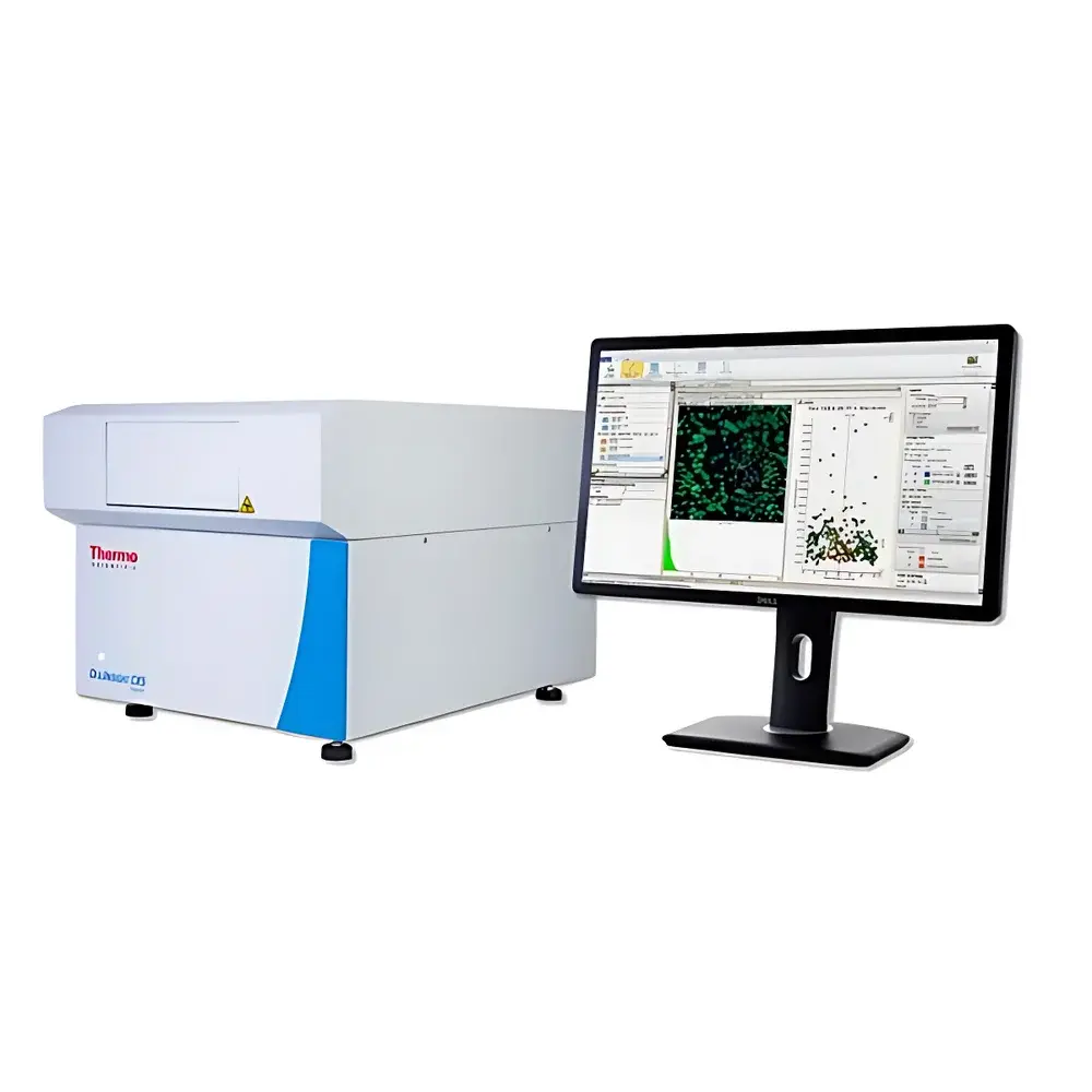
Thermo Scientific CellInsight CX5 High-Content Cell Imaging Analysis System
| Origin | USA |
|---|---|
| Manufacturer Type | Authorized Distributor |
| Origin Category | Imported |
| Model | CellInsight CX5 |
| Price Range | USD 210,000 – 280,000 (based on configuration and regional duties) |
| Temperature Control | Not integrated (requires external environmental chamber for regulated incubation) |
| Humidity Control | Not integrated |
| Cell Analysis Throughput | Up to 500 fields of view per well (96-well plate), ~3–5 min per plate for 4-channel imaging (typical workflow) |
Overview
The Thermo Scientific™ CellInsight™ CX5 High-Content Cell Imaging Analysis System is a compact, benchtop-integrated platform engineered for quantitative, multi-parameter cellular phenotyping in academic, pharmaceutical, and contract research laboratory environments. Based on widefield fluorescence microscopy coupled with high-speed, high-sensitivity sCMOS imaging and precise motorized XYZ stage control, the CX5 enables automated acquisition and algorithm-driven analysis of morphological, spatial, and intensity-based features at single-cell resolution. Unlike conventional endpoint assays or qualitative microscopy, the CX5 implements a rigorous, image-based cytometry paradigm—capturing statistically robust population-level data from thousands of individual cells per well, while preserving spatial context and subcellular localization information. Its design prioritizes reproducibility, walk-away automation, and compliance-ready data handling—making it suitable for early-stage target validation, compound screening, mechanistic toxicology studies, and translational biomarker discovery.
Key Features
- Five-Channel Fluorescence Imaging: Equipped with five LED-based excitation sources and matched emission filters, supporting simultaneous detection of DAPI, FITC, TRITC, Cy5, and far-red probes—enabling co-localization and multiplexed pathway interrogation without spectral overlap artifacts.
- High-Fidelity sCMOS Detection: 2.3 MP scientific CMOS sensor with >73% quantum efficiency, 16-bit dynamic range, and low read noise (<1.5 e⁻) ensures accurate quantification across broad intensity distributions—from dim nuclear markers to saturated cytoplasmic signals.
- Automated Focus & Z-Stack Acquisition: Integrated hardware autofocus (contrast-based) and programmable Z-stack capture (up to 20 planes) support thick samples, organoids, and tissue sections while minimizing focus drift during multi-well runs.
- Benchtop Form Factor with Modular Integration: Compact footprint (54 × 59 × 48 cm) allows deployment in standard biosafety cabinets or shared imaging suites; compatible with third-party environmental chambers (e.g., OKO Labs, INU) for live-cell kinetic assays under controlled CO₂, temperature, and humidity (external control required).
- Pre-Validated Optical Path: Factory-aligned optics with calibrated magnification (10×, 20×, 40× dry objectives), uniform illumination (±3% field flatness), and chromatic aberration correction ensure inter-instrument comparability and longitudinal study consistency.
Sample Compatibility & Compliance
The CellInsight CX5 accommodates standard microplate formats (6–384-well), chambered coverslips, and glass-bottom dishes (24–96 mm). It supports fixed and live-cell preparations—including adherent and suspension cultures, primary neurons, iPSC-derived lineages, 3D spheroids (up to 200 µm diameter), cryosections, and whole-mount zebrafish embryos. All image metadata (acquisition parameters, objective ID, exposure time, gain, binning, date/time stamp) are embedded in TIFF/OME-TIFF files per MIAME and OME-TCF standards. The system complies with FDA 21 CFR Part 11 requirements when deployed with HCS Studio CE-certified software (v3.5+), including electronic signatures, audit trails, user access controls, and immutable raw-data archiving—supporting GLP and GMP-aligned workflows in regulated toxicology and safety pharmacology studies (per OECD TG 487, ICH S7B, USP ).
Software & Data Management
HCS Studio™ Cell Analysis Software (v3.5+) serves as the analytical engine for the CX5 platform. It delivers a modular, workflow-driven interface built on a validated computational framework compliant with ISO/IEC 17025:2017 for measurement uncertainty estimation. Preconfigured analysis modules (n = 32+) cover cell counting, nuclear segmentation, cytoplasmic profiling, neurite tracing, mitotic index scoring, and organelle morphology quantification—each implementing adaptive thresholding, machine-learning-assisted object classification (optional), and batch-processing scalability. Raw image datasets are stored in vendor-neutral OME-TIFF format; processed results export to CSV, Excel, or HDF5 with full traceability to source pixels. Audit logs record every parameter change, algorithm selection, and user action—fully searchable and exportable for regulatory submissions. Integration with Thermo Fisher’s Connect LIMS and third-party platforms (e.g., Dotmatics, Benchling) is supported via RESTful API and secure SFTP protocols.
Applications
- Cell Health & Toxicology: Quantitative assessment of apoptosis (caspase-3/7, Annexin V), necrosis (PI uptake), autophagy (LC3 puncta), ER stress (BiP expression), mitochondrial membrane potential (TMRM), oxidative stress (DCFDA), genotoxicity (γH2AX foci), hepatotoxicity (albumin secretion + steatosis scoring), and neurotoxicity (dendritic arborization loss).
- Oncology & Functional Screening: Phenotypic profiling of migration (wound healing), invasion (Matrigel transwell), adhesion kinetics, proliferation (EdU incorporation), clonogenic survival, angiogenesis (tube formation), and epithelial-mesenchymal transition (E-cadherin/vimentin ratio).
- Signal Transduction Analysis: Subcellular translocation (NF-κB nuclear entry, β-catenin membrane-to-nucleus shift), receptor internalization (EGFR endocytosis), kinase activity reporters (CKAR, FRET biosensors), calcium flux (Fluo-4 kinetics), DNA replication (PCNA foci), and microtubule dynamics (EB3-GFP tracking).
- Stem Cell & Developmental Biology: iPSC differentiation efficiency (SSEA-4/OCT4 co-expression), neural rosette formation, cardiomyocyte beating synchrony (motion vector analysis), myotube fusion index, and zebrafish embryo developmental staging (somite count, heart rate, circulation onset).
FAQ
Does the CellInsight CX5 include integrated temperature and humidity control?
No—the CX5 is a non-incubated platform. For live-cell kinetics, users must integrate an external environmental chamber (e.g., OKO Labs Chamber or PeCon TempModule) with independent CO₂, temperature, and humidity regulation.
Is HCS Studio software qualified for regulated environments?
Yes—HCS Studio CE-certified versions (v3.5 and later) provide 21 CFR Part 11 compliance features, including role-based access control, electronic signatures, and immutable audit trails, validated per IQ/OQ/PQ protocols.
Can the CX5 analyze 3D spheroids or organoids?
Yes—using Z-stack acquisition and extended-depth-of-field reconstruction, the CX5 supports quantitative analysis of spheroids up to 200 µm in diameter, including viability gradients, proliferation zones, and necrotic core detection.
What file formats does the system support for data export?
Raw images: OME-TIFF (metadata-rich, open standard); analysis results: CSV, XLSX, HDF5; reports: PDF with embedded thumbnails and statistical summaries.
How many cells can be analyzed per experiment?
Depending on well density and imaging strategy, typical experiments quantify 1,000–50,000 cells per well—with statistical power validated via bootstrapped confidence intervals and ANOVA-based effect size estimation within HCS Studio.
Related Products


