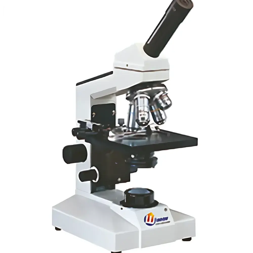
YANRUN BI-12 Biological Inverted Microscope
| Brand | YANRUN |
|---|---|
| Origin | Shanghai, China |
| Manufacturer Type | Direct Manufacturer |
| Product Category | Domestic (China-made) |
| Model | BI-12 |
| Instrument Type | Inverted Microscope |
| Illumination | 20 W Tungsten-Halogen Lamp (110 V / 220 V) or High-Brightness White LED (Optional) |
| Eyepiece | Wide-Field WF10× (Φ18 mm) |
| Objective Lenses | Achromatic 4×/0.10, 10×/0.25, 40×/0.65 (spring-loaded), 100×/1.25 (spring-loaded, oil immersion) |
| Magnification Range | 40×–1000× (standard) |
| Fine Focus Graduation | 4 µm |
| Stage | Fixed mechanical stage (120 mm × 120 mm), travel range 70 mm × 30 mm with vernier scale |
| Nosepiece | Four-position forward-facing ball-bearing turret with external positioning |
| Condenser | Abbe condenser (N.A. 1.25, vertically adjustable) for BI-12B |
| Optional Accessories | WF16× eyepiece, pointer eyepiece, three-position turret, blue/green/yellow filters, 5 W fluorescent lamp, USB/VIDEO trinocular photo-tube, dual-input LED power adapters (9 V / 500 mA) |
Overview
The YANRUN BI-12 Biological Inverted Microscope is an entry-level inverted optical microscope engineered for routine observation of live cells, tissue cultures, and unstained or lightly stained biological specimens in academic teaching labs, quality control environments, and small-scale biotechnology facilities. Its inverted configuration places the objective lenses beneath the specimen stage and the light source above—enabling direct access to culture vessels (e.g., Petri dishes, flasks, and multi-well plates) without disturbing cell monolayers during long-term imaging. The system operates on standard brightfield illumination principles using Köhler illumination geometry, supported by an achromatic objective lens set corrected for chromatic and spherical aberrations across the visible spectrum (400–700 nm). Designed for stability and ergonomic operation, the BI-12 features a rigid cast-metal frame, precision-ground optical pathways, and interchangeable mechanical components to accommodate both educational and light research workflows.
Key Features
- Inverted optical layout optimized for observing adherent cells in standard cultureware—minimizing mechanical disturbance during focus adjustment and stage translation.
- Achromatic objective lens suite (4×, 10×, 40× spring-loaded, 100× oil immersion) delivering consistent resolution and contrast across magnifications from 40× to 1000× (expandable to 1600× with optional WF16× eyepiece).
- Two ergonomic focusing configurations: BI-12A offers separate coarse/fine focus knobs; BI-12B integrates coaxial fine/coarse adjustment with 4 µm fine-focus graduation for precise Z-axis control.
- Fixed mechanical stage (120 mm × 120 mm) with calibrated vernier scales enabling reproducible specimen repositioning over 70 mm × 30 mm travel range.
- Four-position forward-facing nosepiece with ball-bearing indexing ensures repeatable, low-backlash objective alignment and minimizes optical axis shift during lens switching.
- Dual illumination support: factory-configured 20 W tungsten-halogen lamp (110 V / 220 V compatible) or high-luminance white LED module (optional), both compliant with IEC 62471 photobiological safety standards for Class 1 LED devices.
Sample Compatibility & Compliance
The BI-12 accommodates standard transparent-bottom culture vessels—including 35 mm Petri dishes, T25/T75 flasks, and 6-/24-/96-well plates—without requiring coverslip mounting. Its working distance at 40× (≥0.6 mm) and 100× oil (≥0.13 mm) permits unobstructed imaging through common plastic and glass substrates. All optical components meet ISO 10934-1 (Microscopes — Nomenclature of parts) and JIS B 7151 (Japanese Industrial Standard for microscopes). The instrument supports compliance with GLP documentation requirements when used with optional USB/VIDEO output modules for image archiving. While not certified for GMP manufacturing environments per FDA 21 CFR Part 11, its stable mechanical design and traceable calibration path (via included stage micrometer) facilitate audit-ready validation protocols in academic and preclinical settings.
Software & Data Management
The BI-12 does not include proprietary acquisition software but is fully compatible with third-party imaging platforms via its USB/VIDEO trinocular photo-tube option (sold separately). This interface supports real-time streaming to open-source tools such as ImageJ/Fiji, Micro-Manager, and commercial packages including NIS-Elements (Nikon), ZEN (Zeiss), and LAS X (Leica). Video output conforms to NTSC/PAL analog standards; USB variants deliver uncompressed UVC-compliant digital streams at up to 1920×1080@30 fps. Metadata capture—including magnification, objective ID, and timestamp—is enabled when paired with compatible host software. Firmware-free operation ensures deterministic latency and eliminates driver conflicts in multi-instrument lab networks.
Applications
- Routine monitoring of mammalian and insect cell lines (e.g., HeLa, CHO, Sf9) during passage, transfection, and confluence assessment.
- Qualitative evaluation of primary neuron morphology, myoblast fusion, and organoid growth kinetics under ambient CO₂ incubator conditions.
- Teaching laboratory use for introductory histology, microbiology, and developmental biology courses—supporting comparative analysis of stained vs. phase-contrast alternatives.
- QC inspection of sterile filtration membranes, hydrogel scaffolds, and biomaterial coatings where surface topography and cellular adhesion patterns require visual verification.
- Pre-screening prior to advanced modalities (e.g., confocal, time-lapse, or fluorescence imaging) to define ROI selection and exposure parameters.
FAQ
Is the BI-12 suitable for fluorescence imaging?
No—the base model lacks excitation/emission filter sets, mercury/xenon arc lamps, or dichroic mirrors required for epifluorescence. Fluorescence capability requires retrofitting with a dedicated fluorescence attachment kit (not included).
Can the BI-12 be used inside a CO₂ incubator?
No—the instrument is designed for ambient lab use only. It lacks environmental sealing, temperature stabilization, or humidity control necessary for incubator integration.
What is the maximum usable magnification with oil immersion?
At 100× objective and WF10× eyepiece, total magnification is 1000×. With optional WF16× eyepiece, theoretical magnification reaches 1600×, though practical resolution remains limited by the 1.25 NA objective and visible-light wavelength constraints.
Does the BI-12 support phase contrast or darkfield?
Phase contrast and darkfield are not natively supported. These techniques require specialized condensers, annuli, and objectives—none of which are supplied or mechanically compatible with the standard BI-12 optical train.
Is technical documentation available in English?
Yes—full bilingual (English/Chinese) user manuals, mechanical drawings, and optical schematics are provided upon request for regulatory or validation purposes.


