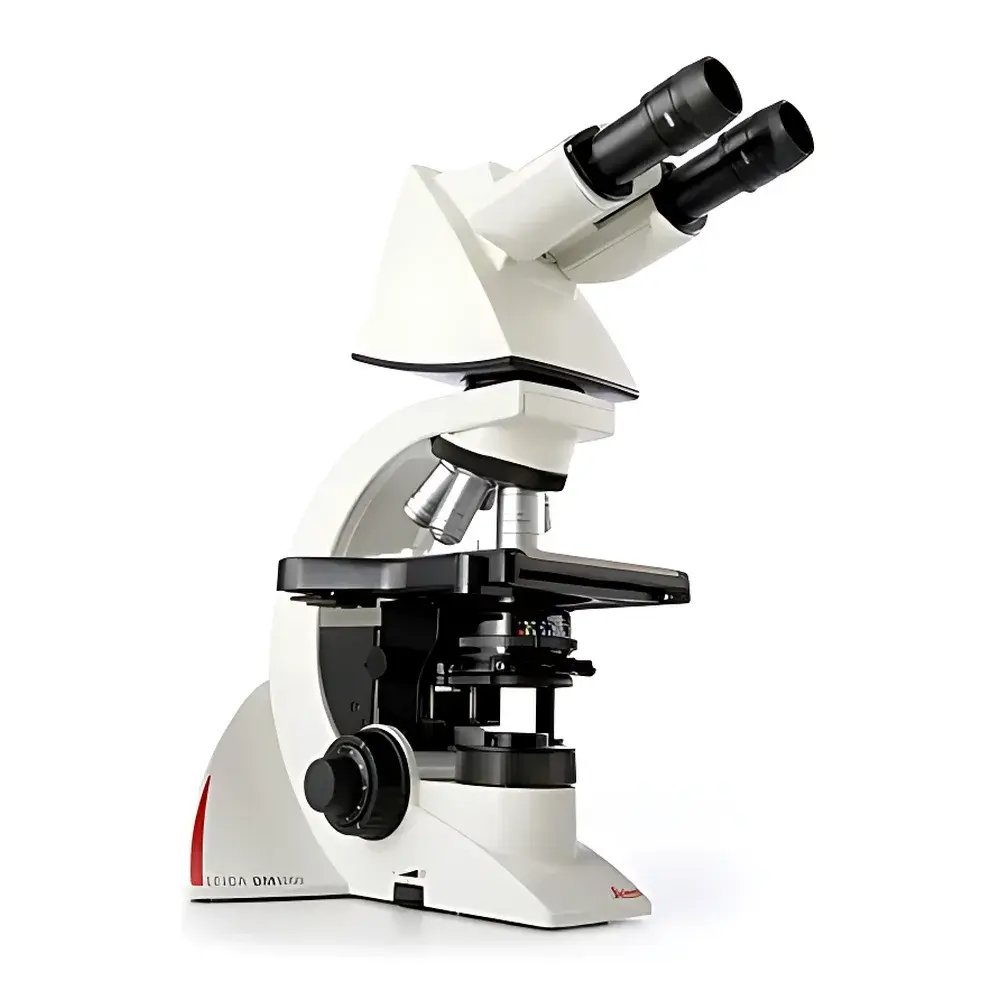
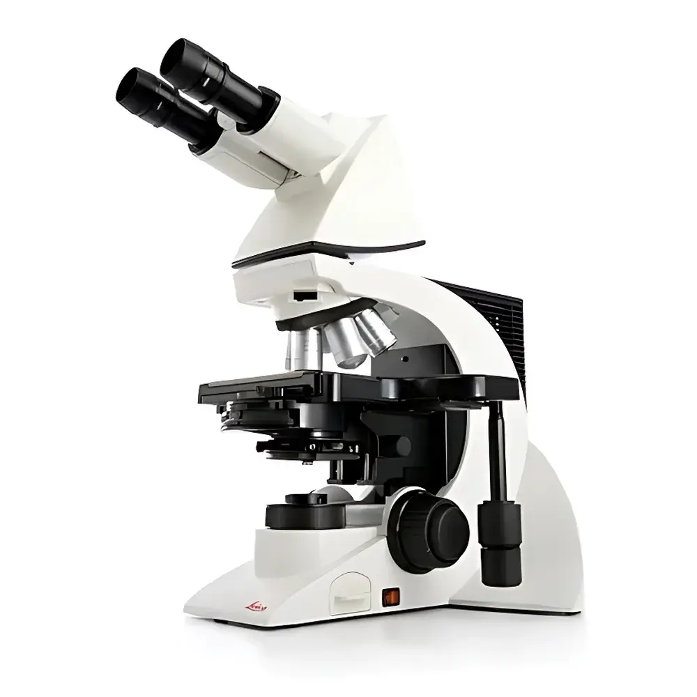

Leica DM1000 / DM2000 / DM2500 Research-Grade Biological Microscope
| Brand | Leica |
|---|---|
| Origin | Germany |
| Model | DM1000 / DM2000 / DM2500 |
| Illumination | LED (DM1000), 100 W Halogen (DM2000/DM2500) |
| Objective Turret | 4–7 position, motorized optional |
| Eyepiece Configuration | Fixed binocular (DM1000), angle-adjustable binocular (DM2000), angle-adjustable trinocular with fluorescence port (DM2500) |
| Fluorescence Capability | Integrated on DM2500 (SFL 100 filter set, excitation @ 470 nm, part no. 11504138) |
| Focus & Stage Control | Ergonomic single-hand coaxial focus + mechanical stage with integrated control (DM1000) |
| Compliance | ISO 9001 certified manufacturing, CE marked, compliant with IEC 61000-6-3 (EMC) and IEC 61000-6-2 (immunity) |
Overview
The Leica DM1000, DM2000, and DM2500 represent a tiered family of research-grade upright biological microscopes engineered for reproducible, high-fidelity optical imaging in academic, clinical, and industrial life science laboratories. All three models are built upon Leica’s modular CTR (Contrast Transfer Ratio) optical platform, featuring apochromatic correction across visible wavelengths and optimized Köhler illumination pathways to ensure uniform intensity, minimal chromatic aberration, and high numerical aperture performance. The DM1000 serves as an entry-level research instrument centered on transmitted-light brightfield and optional darkfield observation, equipped with energy-efficient LED illumination and ergonomic single-hand operation for rapid workflow integration. The DM2000 introduces enhanced contrast management via integrated neutral density and aperture diaphragm controls, a 7-position objective turret, and synchronized coarse/fine focus with mechanical stage translation—ideal for routine histology and cytology screening. The DM2500 constitutes the flagship configuration, integrating a dedicated fluorescence illumination path (100 W HBO lamp or LED alternative), a trinocular head with 20% beam-splitting ratio for simultaneous imaging and observation, and full compatibility with differential interference contrast (DIC) optics—making it suitable for demanding applications in cell dynamics, subcellular localization studies, and dual-modal (brightfield + fluorescence) correlative analysis.
Key Features
- Ergonomic optical design: All models feature low-positioned focus knobs, adjustable interpupillary distance, and height-variable eyepiece tubes to reduce operator fatigue during extended use.
- Modular contrast enhancement: DM2000 and DM2500 support phase contrast (Ph1–Ph4), darkfield (DF), and polarized light accessories; DM2500 adds DIC capability with strain-free objectives and Nomarski prisms.
- Fluorescence-ready architecture: DM2500 includes a dedicated fluorescence port, SFL 100 filter cube (excitation 470 ± 20 nm, emission 525 ± 25 nm, dichroic 500 nm), and optional motorized filter changer for multi-channel acquisition.
- Precision mechanical stage: Integrated X-Y translation with vernier scales (0.1 mm resolution), specimen holder clips, and optional encoding for coordinate tracking compatible with Leica Application Suite (LAS X) software.
- Thermally stable illumination: DM2000/DM2500 utilize stabilized 100 W halogen lamps with IR-filtered condensers; DM1000 employs constant-current LED source (6,000 K CCT) with >50,000-hour lifetime and no warm-up delay.
Sample Compatibility & Compliance
The DM-series accommodates standard glass microscope slides (26 × 76 mm), petri dishes (35–100 mm), multi-well plates (6–96-well), and live-cell chambers (e.g., Lab-Tek chambered coverglass). All models comply with ISO 10993-5 (biocompatibility of optical components contacting specimens), IEC 61000-6-3 (EMC emissions), and IEC 61000-6-2 (electromagnetic immunity). The DM2500 meets additional requirements for clinical pathology environments per CLSI EP17-A2 (interference testing) and supports audit-trail-enabled operation when paired with LAS X software under 21 CFR Part 11-compliant configurations (user authentication, electronic signatures, change logs). Optical components adhere to DIN EN ISO 10110-7 surface quality standards (scratch-dig 20–10), and all objectives are sealed against dust and humidity per IP54 rating.
Software & Data Management
Leica DM microscopes integrate natively with Leica Application Suite (LAS X) v3.7+, supporting image acquisition, annotation, measurement (area, length, intensity profiling), Z-stack reconstruction, and time-lapse registration. LAS X provides GLP/GMP-compliant metadata embedding (operator ID, timestamp, objective magnification, illumination settings, exposure parameters) and export to TIFF, OME-TIFF, and JPEG2000 formats. For DM2500 users performing quantitative fluorescence, LAS X includes background subtraction algorithms, spectral unmixing (with multi-band filter sets), and ROI-based intensity normalization referenced to internal calibration slides. Data integrity is reinforced through encrypted local storage, role-based access control, and optional network deployment with centralized license management.
Applications
- Histopathology: Routine H&E, PAS, and trichrome staining evaluation with high-resolution 40× and 63× oil-immersion objectives.
- Hematology: Peripheral blood smear analysis, reticulocyte counting, and blast cell identification using phase contrast and brightfield modes.
- Cell culture monitoring: Live/dead assessment via calcein-AM/ethidium homodimer staining on DM2500 with temperature-controlled stage adapters.
- Microbiology: Gram staining interpretation, fungal morphology, and motility analysis using darkfield condensers and 100× oil objectives.
- Academic research: Co-localization studies (e.g., GFP/RFP dual labeling), mitotic staging, and organelle dynamics tracking with time-lapse fluorescence modules.
FAQ
Is the DM1000 compatible with fluorescence imaging?
No—the DM1000 lacks a fluorescence illumination path, filter cube housing, and appropriate dichroic mirrors. Fluorescence capability is exclusive to the DM2500 configuration.
Can the DM2000 be upgraded to support DIC?
Yes—provided the base model includes a strain-free condenser and matched DIC-compatible objectives (e.g., HC PL APO 40×/0.85 or 63×/1.32), a DIC slider and prism set can be retrofitted via Leica Service Center calibration.
What is the maximum working distance for oil-immersion objectives on the DM2500?
The standard HC PL FLUOTAR 100×/1.30 Oil objective offers a working distance of 0.15 mm; optional long-working-distance variants (e.g., HC PL APO 63×/1.40 CORR) support coverslip thickness correction from 0.13–0.21 mm.
Does LAS X software support automated cell counting?
Yes—LAS X includes a trainable machine-learning module (LAS X Count & Measure) that supports binary thresholding, size/gating filters, and classification based on morphological features (circularity, aspect ratio, texture).
Are replacement parts and service documentation available globally?
Yes—Leica Microsystems maintains authorized service centers in over 40 countries, with online access to technical bulletins, calibration certificates, and RoHS/REACH compliance declarations via the Leica Support Portal.


