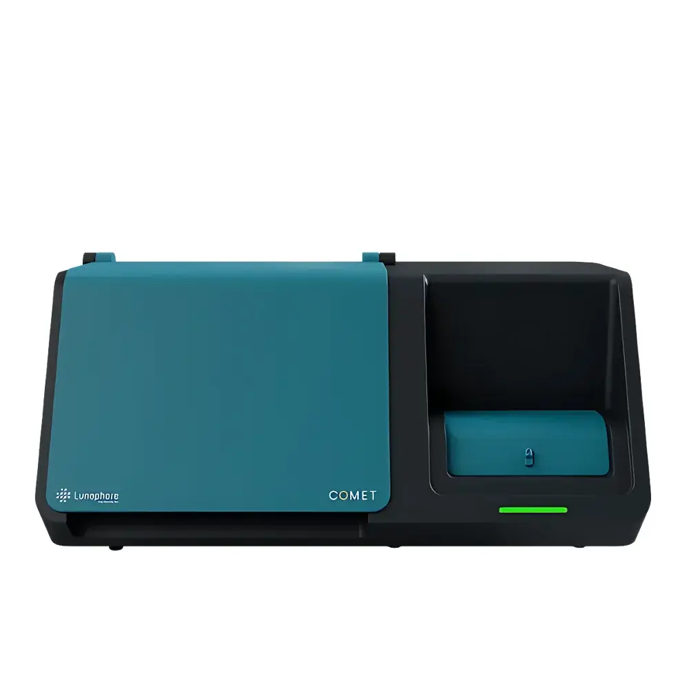
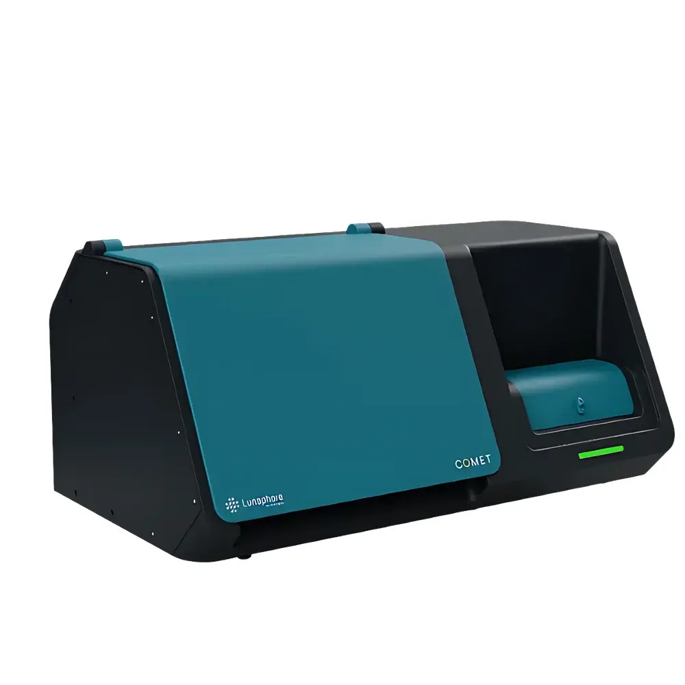
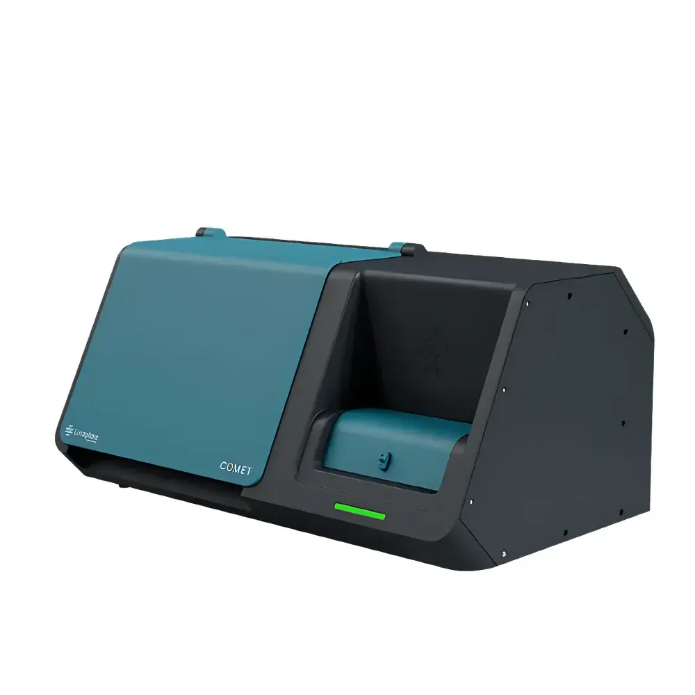
Bio-Techne COMET™ Automated Single-Cell Spatial Multi-Omics Platform
| Brand | Bio-Techne |
|---|---|
| Origin | Switzerland |
| Manufacturer Type | Authorized Distributor |
| Origin Category | Imported |
| Model | COMET |
| Automation Level | Fully Automated |
| Instrument Type | Fluorescence In Situ Hybridization (FISH) & Sequential Immunofluorescence (seqIF™) Platform |
| Sample Capacity | 4 Slides per Run |
| Compatible Sample Types | FFPE and Fresh-Frozen Tissue Sections |
| Core Technology | seqIF™ (Sequential Immunofluorescence) + RNAscope™ HiPlex Pro In Situ Hybridization |
| Max Protein Targets per Run | 40-plex |
| Max RNA Targets per Run | 12-plex |
| Integrated Workflow | Automated Staining, Hybridization, Imaging, Antibody Stripping, and OME-TIFF Output |
Overview
The Bio-Techne COMET™ Automated Single-Cell Spatial Multi-Omics Platform is an engineered solution for high-fidelity, subcellular-resolution spatial profiling of both RNA and protein within intact tissue architecture. Built upon Lunaphore’s proprietary sequential immunofluorescence (seqIF™) and RNAscope™ HiPlex Pro in situ hybridization technologies, COMET™ delivers fully automated, iterative cycles of antibody staining, fluorescence imaging, and gentle antibody elution—without covalent antibody conjugation or barcode labeling. This eliminates the need for custom-labeled reagents while preserving epitope integrity and tissue morphology across multiple rounds. The platform operates on standard glass slides containing formalin-fixed paraffin-embedded (FFPE) or cryopreserved tissue sections, enabling reproducible, quantitative spatial phenotyping at single-cell resolution. Its closed-loop architecture supports end-to-end automation—from probe hybridization and signal amplification to multi-channel image acquisition and digital registration—resulting in a unified OME-TIFF dataset that integrates spatial transcriptomic and proteomic information from the same physical section.
Key Features
- Fully automated, walk-away operation for sequential immunofluorescence (seqIF™) and RNAscope™ HiPlex Pro workflows
- 4-slide capacity per run with independent protocol scheduling and temperature-controlled incubation zones
- Subcellular resolution imaging across up to 12 RNA targets and 40 protein targets on a single tissue section
- Compatibility with off-the-shelf, unconjugated primary antibodies and commercially available fluorophore-conjugated secondary antibodies
- Gentle, buffer-based antibody stripping that maintains tissue morphology and enables downstream validation (e.g., H&E, IHC, ISH)
- Integrated DAPI nuclear counterstaining and cross-cycle image alignment for precise spatial registration
- Modular reagent reservoirs supporting extended multiplexing beyond 40-plex via reloaded cycles without slide removal
Sample Compatibility & Compliance
COMET™ is validated for human and murine FFPE and frozen tissue sections—including delicate specimens such as bone marrow, skin, lung, and pancreas—and demonstrates robust performance across diverse species including rat, canine, and non-human primate tissues. All protocols adhere to Good Laboratory Practice (GLP) principles and support audit-ready documentation. The platform’s reagent handling, temperature calibration logs, and imaging metadata comply with FAIR data standards and are compatible with 21 CFR Part 11–compliant LIMS integration when deployed in regulated environments. While not a diagnostic device, COMET™ workflows align with ISO 15189 pre-analytical requirements for tissue-based biomarker assays and support method validation per CLSI EP17-A2 guidelines for low-abundance target detection.
Software & Data Management
The COMET™ system runs on embedded firmware synchronized with HORIZON™ software—a dedicated image analysis suite designed for spatial multi-omics quantification. HORIZON™ provides automated nuclear segmentation using pretrained deep learning models (U-Net architecture), cell boundary refinement via marker-guided watershed, and pixel-level registration of sequential RNA and protein channels. All raw and processed data—including TIFF stacks, alignment matrices, and feature tables—are exported in OME-TIFF format compliant with the Open Microscopy Environment specification. Metadata embedding follows MIAME and MINSEQE standards, facilitating integration with downstream tools such as Seurat, Squidpy, or Visium-compatible pipelines. Audit trails record every instrument action—including reagent lot numbers, exposure times, focus offsets, and elution cycle counts—for full traceability.
Applications
- Spatially resolved tumor microenvironment mapping across immune cell subsets, stromal components, and malignant clones
- Validation of scRNA-seq-derived biomarkers in native tissue context with concurrent protein expression confirmation
- Mechanistic studies of post-transcriptional regulation through direct comparison of RNA abundance and corresponding protein localization
- Longitudinal biomarker discovery in clinical trial biobanks using archival FFPE cohorts
- Development of spatially informed predictive signatures for immuno-oncology response assessment
- Neurodegenerative disease research requiring co-detection of misfolded proteins and associated transcript isoforms
FAQ
Does COMET™ require custom-conjugated antibodies?
No. COMET™ uses standard unconjugated primary antibodies and commercial fluorophore-conjugated secondary antibodies—eliminating time-consuming conjugation steps and ensuring compatibility with existing lab inventories.
Can tissue be reused after COMET™ processing?
Yes. The mild elution chemistry preserves tissue morphology and antigenicity, allowing subsequent H&E staining, IHC, or additional ISH assays on the same slide.
What is the maximum multiplexing capability?
A single run supports up to 40 protein targets and 12 RNA targets. Additional cycles can be initiated by reloading reagents, enabling theoretically unlimited plexing on one section.
Is RNAscope™ HiPlex Pro included or licensed separately?
RNAscope™ HiPlex Pro probes and assay kits are supplied under license from Advanced Cell Diagnostics (a Bio-Techne company); probe design and ordering are managed through the ACD portal.
How is image registration maintained across sequential cycles?
DAPI-based nuclear landmarks and affine transformation algorithms ensure sub-pixel alignment between all RNA and protein imaging rounds, with residual registration error < 0.3 µm RMS.


