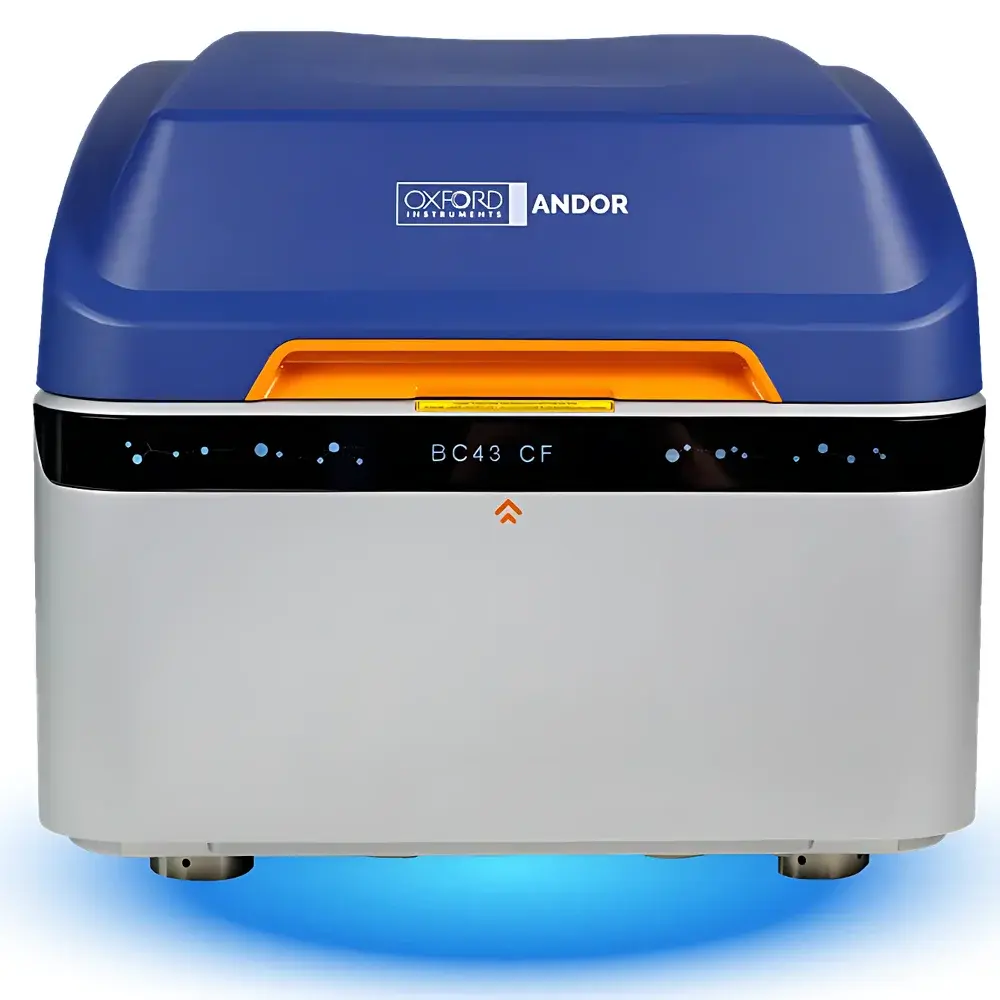
Andor BC43 SR Desktop Super-Resolution Confocal Microscope
| Brand | Oxford Instruments |
|---|---|
| Origin | United Kingdom |
| Model | BC43 SR |
| Pixel Size | 6.5 µm |
| Resolution | 2040 × 1992 (4.1 MP) |
| Excitation Lasers | 405 nm, 488 nm, 561 nm, 639 nm |
| Scanning Mode | XYZ |
| Objective Lenses | 10×, 20×, 40×, 60×, 100× |
| Software & Workstation | Imaris image analysis software |
| Vibration Isolation | Integrated active/passive damping platform |
| XY Stage Control | Motorized |
Overview
The Andor BC43 SR is a compact, benchtop super-resolution confocal microscope engineered for high-fidelity fluorescence imaging in life science laboratories where space, workflow efficiency, and quantitative reproducibility are critical. Built upon the proven optical architecture of Andor’s award-winning Dragonfly platform, the BC43 SR implements spinning-disk confocal technology combined with structured illumination or deconvolution-enhanced super-resolution modalities—depending on configuration—to achieve effective lateral resolution beyond the diffraction limit (~120–180 nm). Unlike traditional upright or inverted research-grade confocal systems requiring dedicated darkrooms and complex alignment, the BC43 SR operates reliably on standard laboratory benches, eliminating infrastructure overhead while maintaining rigorous optical performance. Its integrated laser combiner supports four solid-state excitation wavelengths (405/488/561/639 nm), enabling multicolor immunofluorescence, live-cell Ca²⁺ imaging, FRET, and organelle dynamics studies—all without manual filter wheel changes. The system adheres to ISO 19012-1 (microscope terminology) and complies with CE marking requirements for in vitro diagnostic (IVD) and research-use-only (RUO) instrumentation.
Key Features
- Compact benchtop form factor with integrated vibration-damped optical baseplate—no external anti-vibration table required
- Motorized XYZ scanning stage with sub-micron repeatability and programmable multi-position acquisition
- High-sensitivity sCMOS detection optimized for low-light confocal and super-resolution modes
- Pre-aligned, field-serviceable laser launch module with TTL-triggered wavelength switching
- Standard objective turret accommodating 10× to 100× oil/water/glycerol immersion objectives (Nikon CFI Plan Apo or equivalent)
- Factory-calibrated IQ/OQ validation protocol per ISO/IEC 17025 guidelines, including intensity uniformity, spatial resolution verification, and Z-drift characterization
Sample Compatibility & Compliance
The BC43 SR accommodates standard glass-bottom dishes (35 mm, 60 mm), multi-well plates (24-, 48-, 96-, and 384-well), and custom-mounted tissue sections up to 1 mm thickness. It supports live-cell imaging at ambient temperature or with optional environmental chamber integration (CO₂, humidity, temperature control). All optical components comply with RoHS 2011/65/EU and REACH (EC 1907/2006) material restrictions. For regulated environments, the system supports audit-ready metadata logging—including timestamped acquisition parameters, user ID, objective magnification, laser power settings, and detector gain—enabling alignment with GLP/GMP documentation workflows and FDA 21 CFR Part 11 electronic record requirements when paired with validated Imaris deployment.
Software & Data Management
Imaris software serves as the native acquisition and analysis engine, delivering real-time 3D rendering, automated spot/ filament/surface segmentation, and batch-processing pipelines compatible with FIJI/ImageJ macro scripting. Acquisition modules include time-lapse (4D), Z-stack (5D), multi-channel tiling, and spectral unmixing. Raw data is saved in standardized OME-TIFF format with embedded metadata compliant with the Open Microscopy Environment (OME) schema. The included workstation (4 TB or 8 TB RAID 5 configuration) ensures local high-throughput storage with hardware-accelerated compression (JPEG2000 or lossless LZ4). Optional cloud synchronization via secure SFTP or DICOM-SR export enables cross-institutional collaboration and long-term archival per NIH Data Management and Sharing Policy (DMS) standards.
Applications
- Subcellular localization and co-localization analysis of endogenous or tagged proteins in fixed and live mammalian cells
- Quantitative assessment of nuclear morphology, mitochondrial network dynamics, and lysosomal pH gradients
- 3D reconstruction of thick tissue sections (e.g., brain slices, tumor biopsies) using optical sectioning depth up to 100 µm
- Time-resolved tracking of vesicle trafficking, mitotic spindle assembly, and cytokinetic ring formation
- Validation of CRISPR-edited cell lines via morphometric phenotyping and intensity-based reporter quantification
- Teaching laboratory implementation—pre-configured protocols reduce setup time and minimize user-dependent variability
FAQ
Is the BC43 SR suitable for live-cell imaging over extended durations?
Yes—the system features low-phototoxicity spinning-disk confocal illumination, thermally stabilized detectors, and optional environmental control modules to maintain physiological conditions during multi-hour acquisitions.
Can I upgrade from widefield (BC43 WF) or standard confocal (BC43 CF) to super-resolution (BC43 SR) post-purchase?
Yes—hardware and software upgrades are supported through Andor-certified field service engineers, subject to optical path compatibility and detector firmware revision.
Does the system support third-party objective lenses?
It accepts standard RMS-threaded or Nikon CFI-mount objectives; however, optimal performance (especially for super-resolution modes) requires use of Andor-validated lenses with specified back focal plane and transmission profiles.
What level of technical support and warranty is provided?
All BC43 SR systems ship with a 24-month comprehensive warranty covering parts, labor, and on-site IQ/OQ revalidation; extended service plans include priority remote diagnostics and annual performance verification.
How does the BC43 SR ensure data traceability for publication or regulatory submission?
Every acquired image embeds EXIF-like metadata (including laser power, exposure time, gain, objective ID, and calibration timestamps); Imaris export modules generate PDF reports with embedded audit trails compliant with journal requirements (e.g., Nature Methods reporting standards).


