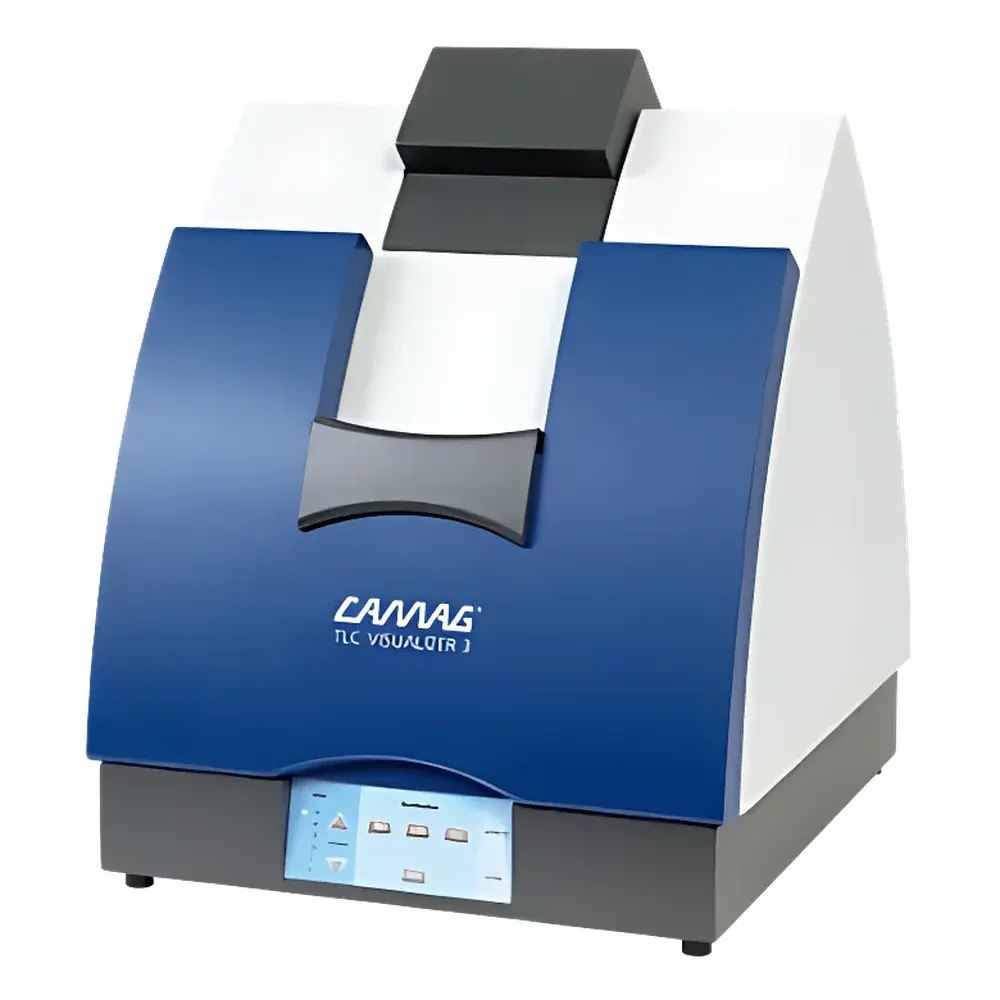
CAMAG TLC Visualizer 3 Digital Imaging System for Thin-Layer Chromatography
| Brand | CAMAG |
|---|---|
| Origin | Switzerland |
| Model | TLC Visualizer 3 |
| Light Sources | 254 nm short-wave UV (epi-illumination), 366 nm long-wave UV (epi-illumination), white light (epi-illumination and trans-illumination) |
| Camera Sensor | High-resolution industrial CMOS |
| Software Platform | visionCATS v3.x or later |
| Compliance | Designed for GLP-compliant laboratories |
Overview
The CAMAG TLC Visualizer 3 is a dedicated digital imaging system engineered for standardized documentation, quantitative evaluation, and archival of thin-layer chromatography (TLC) and high-performance thin-layer chromatography (HPTLC) plates. It operates on the principle of controlled epi- and trans-illumination combined with high-fidelity digital capture, enabling reproducible visualization of analyte zones under multiple spectral conditions—critical for method development, identity confirmation, purity assessment, and stability-indicating analysis. Unlike general-purpose document scanners or consumer-grade cameras, the TLC Visualizer 3 integrates optical path calibration, uniform illumination geometry, and plate-specific positioning fixtures to eliminate spatial distortion and intensity variability across repeated acquisitions. Its architecture conforms to the physical constraints of HPTLC plates (e.g., 10 × 10 cm, 10 × 20 cm, 20 × 20 cm), ensuring full-plate coverage without manual stitching or edge interpolation.
Key Features
- Industrial-grade CMOS camera with fixed focal length lens and motorized focus adjustment, optimized for consistent depth-of-field across standard HPTLC plate thicknesses (0.1–0.25 mm silica layers).
- Dual-mode illumination system: short-wave UV (254 nm) and long-wave UV (366 nm) lamps provide epi-illumination only; white light source supports both epi- and trans-illumination—enabling detection of UV-active, fluorescent, and non-fluorescent compounds via derivatization or inherent chromophores.
- Integrated plate stage with mechanical alignment guides and pressure-free clamping, minimizing plate deformation during imaging and preserving Rf integrity.
- visionCATS software suite (v3.5+), compliant with FDA 21 CFR Part 11, featuring role-based user permissions, electronic signature workflows, and tamper-evident audit trails for all image acquisition, processing, and reporting events.
- Automated exposure time optimization and real-time histogram preview, reducing operator dependency and enhancing inter-day reproducibility.
Sample Compatibility & Compliance
The TLC Visualizer 3 accommodates all commercially available HPTLC plates—including silica gel 60 F254, aluminum-backed cellulose, polyamide, and reversed-phase C18—regardless of backing material (glass, aluminum, or polyester). It supports post-chromatographic derivatization workflows (e.g., anisaldehyde/H2SO4, ninhydrin, Dragendorff’s reagent) followed by immediate imaging under white light or UV. The system meets ISO/IEC 17025 requirements for testing laboratories when operated within validated SOPs and calibrated using CAMAG-certified reference plates. All firmware and software updates are traceable through CAMAG’s GxP-aligned release documentation, supporting regulatory inspections under EU Annex 11 and ICH Q5C guidelines.
Software & Data Management
visionCATS provides native support for multi-wavelength overlay, Rf calculation, spot integration (peak area/height), and calibration curve generation using internal standards or external references. Image enhancement tools—including “Spot Magnification” (digital zoom with pixel interpolation correction), “White Reference Correction” (background subtraction using blank plate images), and “Exposure Normalization” (intensity scaling across batches)—are algorithmically validated and fully auditable. Processed data exports to CSV, PDF/A-2, TIFF (64-bit), and vendor-neutral XML formats compatible with LIMS and ELN systems. Raw image files retain EXIF metadata (exposure time, lamp status, plate ID, timestamp), ensuring full traceability from acquisition to report.
Applications
- Pharmaceutical quality control: identification and assay of active pharmaceutical ingredients (APIs) and impurities per USP and Ph. Eur. 2.2.27.
- Natural product fingerprinting: comparative analysis of herbal extracts across seasonal harvests or extraction methods.
- Food safety screening: detection of mycotoxins (aflatoxins, ochratoxin A), pesticides, and adulterants using multi-step derivatization protocols.
- Forensic toxicology: rapid profiling of drugs of abuse in biological matrices following solid-phase extraction and HPTLC separation.
- Academic research: method validation studies requiring documented repeatability (RSD <3% for Rf and peak area) and inter-laboratory transferability.
FAQ
Does the TLC Visualizer 3 support fluorescence quantification?
Yes—when used with 366 nm epi-illumination and appropriate fluorescent plates or post-derivatization, the system captures linear-intensity response over 3 orders of magnitude (verified with quinine sulfate standards).
Can images be acquired in compliance with 21 CFR Part 11?
Yes—visionCATS implements full electronic signature capability, audit trail logging, and user-level access controls, provided the host computer environment meets NIST SP 800-53 security baselines.
Is trans-illumination available for all plate types?
Trans-illumination is supported only for transparent-backing plates (e.g., glass or polyester); aluminum-backed plates require epi-illumination exclusively.
What is the minimum detectable spot size under UV 254 nm?
With standard silica gel 60 F254 plates and 100 ng loading, the system resolves discrete spots ≥0.5 mm in diameter at S/N >10.
How often does the system require recalibration?
No routine recalibration is needed; however, annual verification using CAMAG’s Traceable Reference Plate Kit (Order No. 20.0101) is recommended for GLP environments.
Related Products


")