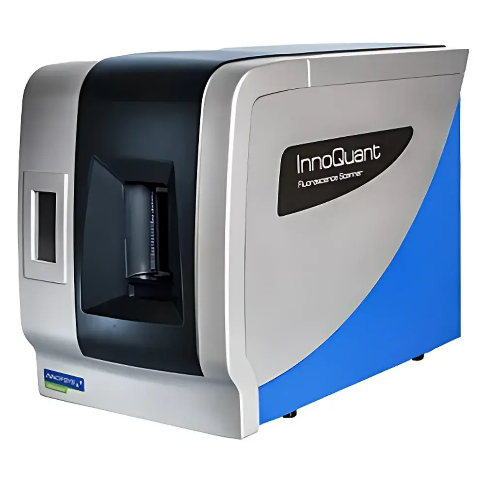
InnoQuant Laser Confocal Fluorescence Quantitative Tissue Slide & Biochip Scanner
| Brand | INNOPSYS |
|---|---|
| Origin | Germany |
| Model | InnoQuant |
| Laser Wavelengths | 375 nm, 488 nm, 561 nm, 640 nm |
| Detection Channels | 4 independent PMT detectors |
| Filter Wheels | 7-position per channel |
| Spatial Resolution | 0.5 µm/pixel |
| Scan Area | 18 mm × 18 mm (full slide, no tiling) |
| Scan Time | ≤17 min @ 0.5 µm/pixel |
| Output Format | 8-bit or 16-bit TIFF |
| Imaging Mode | Point-scanning confocal |
| Z-axis Auto-focus | Real-time dynamic focus tracking |
Overview
The InnoQuant Laser Confocal Fluorescence Quantitative Tissue Slide & Biochip Scanner is a high-precision, research-grade imaging platform engineered for quantitative fluorescence analysis of histological sections, cytological preparations, and microarray-based biochips. Designed and manufactured in Germany by INNOPSYS, the system employs true point-scanning laser confocal architecture—distinct from widefield or spinning-disk approaches—to eliminate out-of-focus blur, suppress autofluorescence background, and deliver optical sectioning capability with sub-micron spatial fidelity. Its four solid-state lasers (375 nm, 488 nm, 561 nm, 640 nm) span the UV-to-red spectral range, enabling excitation of common fluorophores including DAPI, FITC, TRITC, Cy5, and newer near-infrared dyes. Each laser is coupled to an independent photomultiplier tube (PMT) detector with real-time gain and offset control, ensuring linear signal response across dynamic ranges exceeding 4 orders of magnitude. The system operates without field-of-view tiling or image stitching, acquiring full-slide (18 mm × 18 mm) data in a single continuous scan—preserving pixel-level intensity integrity and eliminating edge artifacts inherent in mosaic-based digital pathology systems.
Key Features
- Four independently addressable laser sources with precise wavelength stability and low-noise output for multiplexed excitation of diverse fluorophore panels
- Four synchronized PMT detectors, each equipped with a motorized 7-position filter wheel for flexible emission band selection and optimal spectral separation
- True confocal point-scanning optics delivering isotropic resolution of 0.5 µm/pixel—validated per ISO 19012-1 for lateral resolution calibration
- Real-time Z-axis dynamic autofocus using reflected laser signal feedback, eliminating manual refocusing and enabling consistent focus across uneven tissue surfaces
- Full-scan acquisition mode: no tile-based acquisition, no software-based image stitching, no intensity normalization post-processing—ensuring raw pixel intensity values are directly proportional to local fluorophore concentration
- Native support for both 8-bit and 16-bit TIFF export with embedded metadata (including laser power, PMT gain, dwell time, pinhole size, and objective magnification)
Sample Compatibility & Compliance
The InnoQuant accommodates standard glass microscope slides (1″ × 3″, up to 1.2 mm thickness), including charged, poly-L-lysine-coated, and silanized substrates used for tissue microarrays (TMAs), FFPE sections, frozen sections, cytospin preparations, and spotted nucleic acid or protein biochips. It supports conventional mounting media (e.g., DPX, Vectashield) and aqueous-based antifade reagents. All optical components comply with IEC 60825-1:2014 Class 3B laser safety standards, and the instrument enclosure meets EN 61000-6-3 EMC emission requirements. Data acquisition workflows are compatible with GLP/GMP environments: audit trail logging, user access controls, and electronic signature support can be enabled via optional software modules aligned with FDA 21 CFR Part 11 requirements.
Software & Data Management
Control and analysis are performed via INNOPSYS’ proprietary InnoScan Suite v5.x, a Windows-based application developed in compliance with ISO/IEC 17025 software validation guidelines. The suite provides non-destructive raw data handling, batch processing pipelines for multi-slide experiments, and integrated tools for region-of-interest (ROI) definition, cell segmentation (via intensity-threshold and shape-based algorithms), and spatial statistics (e.g., nearest-neighbor distance, co-localization coefficients). Export formats include OME-TIFF for interoperability with QuPath, ImageJ/Fiji, HALO, and Visiopharm platforms. All acquired datasets retain embedded EXIF-like metadata compliant with MIAME and MIAPE reporting standards for reproducible computational analysis.
Applications
- Digital pathology: High-fidelity whole-slide scanning of H&E and multiplex immunofluorescence (mIF) stained tissue sections for biomarker quantification and spatial phenotyping
- Translational research: Quantitative comparison of protein expression gradients across tumor-stroma interfaces in FFPE cohorts
- Plant and animal histology: Subcellular localization studies in thick-sectioned botanical samples or embryonic tissue stacks
- Biochip analysis: Fluorescence intensity mapping of DNA methylation arrays, SNP genotyping chips, or antibody microarrays with calibrated signal linearity
- Cytology digitization: Objective, reproducible scoring of fluorescently labeled cervical smears or bone marrow aspirates under standardized illumination conditions
- Long-term archival: Generation of stable, vendor-neutral TIFF archives suitable for institutional biobank integration and federated data sharing initiatives
FAQ
Does the InnoQuant require periodic optical recalibration?
Yes—INNOPSYS recommends annual factory calibration using NIST-traceable fluorescence reference standards (e.g., Chroma Tech QF-100 series) to maintain quantitative accuracy across laser lines and PMT channels.
Can the system be integrated into existing LIMS or pathology information systems (PIS)?
Yes—the InnoScan Suite supports DICOM-SR export and offers a RESTful API for bidirectional communication with laboratory middleware, enabling automated job queuing and metadata synchronization.
Is Z-stack acquisition supported?
Yes—users may define custom Z-intervals (0.1–10 µm steps) and acquire serial optical sections; maximum stack depth is limited only by sample transparency and signal-to-noise ratio at depth.
What objective lenses are compatible?
The system ships with a motorized 20×/0.75 NA air objective optimized for fluorescence; optional 10×, 40×, and 63× oil-immersion objectives are available with matched tube lens configuration and automatic magnification detection.
How is photobleaching minimized during acquisition?
By combining low-dwell-time point scanning (≤10 µs/pixel), adjustable laser power per channel (0.1–100% in 0.1% increments), and real-time PMT gain optimization, the InnoQuant reduces total photon dose by up to 60% compared to widefield CCD-based scanners performing equivalent resolution scans.


