68096 Mouse Spinal Cord Experimental Setup
| Origin | Spain |
|---|---|
| Manufacturer Type | Authorized Distributor |
| Origin Category | Imported |
| Model | 68096 |
| Pricing | Available Upon Request |
Overview
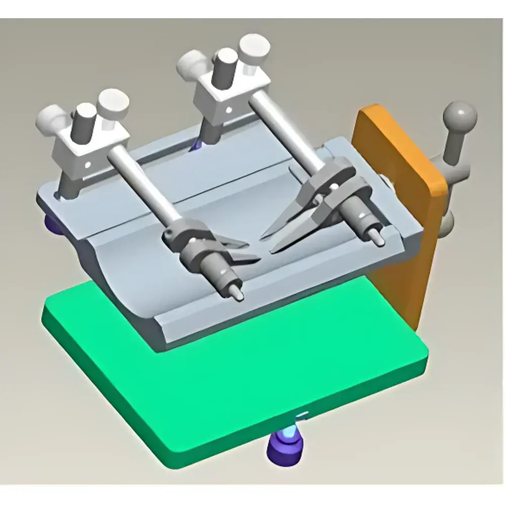
The 68096 Mouse Spinal Cord Experimental Setup is a precision-engineered stereotaxic accessory designed specifically for in vivo spinal cord manipulation and imaging in murine models. Unlike conventional restraint systems, this device employs a gravity-neutral suspension configuration that isolates the spinal column from thoracic and diaphragmatic motion artifacts—critical for high-resolution intravital microscopy, electrophysiological recording, or targeted microinjection into the lumbar or thoracic spinal cord. Its mechanical architecture is grounded in stereotaxic principles adapted from cranial neurosurgery instrumentation but reconfigured to accommodate the anatomical constraints and biomechanical requirements of rodent spinal positioning. The system does not incorporate motorized actuation or integrated sensors; instead, it delivers passive, repeatable, and operator-controlled spatial stabilization—ensuring compatibility with standard upright or inverted research-grade microscopes equipped with motorized stages and long-working-distance objectives.
Key Features
- Gravity-compensated dorsal suspension design minimizes respiratory-induced movement artifacts during real-time imaging or electrophysiology.
- Bi-axial rotational capability (left/right yaw) with angle modulation governed by adjustable support plate height—enabling optimal spinal exposure across L1–L6 vertebral levels.
- T-slot compatible base with integrated T-bolt fastening system for rigid, vibration-damped attachment to standard microscope stage plates (e.g., Leica, Zeiss, Nikon, Olympus).
- Modular aluminum alloy construction ensures dimensional stability under thermal drift conditions typical in extended-duration experiments (≥2 hr).
- Non-invasive clamping interface with silicone-coated contact surfaces prevents tissue compression while maintaining positional fidelity throughout experimental protocols.
Sample Compatibility & Compliance
The 68096 setup is validated for use with C57BL/6, BALB/c, and CD-1 adult mice (20–30 g body weight). It supports both acute terminal preparations and survival surgeries when combined with appropriate anesthesia and postoperative care protocols. All materials comply with ISO 10993-5 (biological evaluation of medical devices — cytotoxicity) and EU Directive 2001/95/EC on general product safety. While the device itself is not classified as a medical device under FDA 21 CFR Part 820 or MDR 2017/745, its application in preclinical neuroscience research aligns with AAALAC International accreditation standards for animal model instrumentation. Documentation includes CE marking per Directive 2014/30/EU (EMC) and 2014/35/EU (LVD), confirming electromagnetic compatibility and low-voltage safety for laboratory environments.
Software & Data Management
This is a hardware-only mechanical platform with no embedded firmware, onboard memory, or proprietary software interface. It operates independently of acquisition systems and integrates seamlessly with third-party imaging platforms—including NIS-Elements (Nikon), ZEN (Zeiss), LAS X (Leica), and MicroManager—via standard mechanical synchronization triggers (TTL pulse input/output). All positional configurations are manually recorded using calibrated micrometer dials and documented within electronic lab notebooks (ELNs) compliant with 21 CFR Part 11 audit trail requirements. Users are advised to log setup parameters—including plate height, rotation angle, and vertebral level alignment—in metadata fields associated with corresponding image stacks or electrophysiology recordings.
Applications
- Intravital two-photon imaging of spinal cord microvasculature, immune cell trafficking, or neuronal calcium dynamics.
- Microelectrode array (MEA) or patch-clamp recordings from dorsal horn neurons during sensory stimulation paradigms.
- Image-guided intraparenchymal injection of viral vectors, tracers, or therapeutics into defined spinal laminae.
- Ex vivo spinal cord slice preparation where precise dorsoventral orientation must be preserved prior to sectioning.
- Validation studies requiring reproducible inter-animal spinal alignment for comparative histomorphometry or immunohistochemical quantification.
FAQ
Is the 68096 compatible with stereotaxic frames from other manufacturers?
Yes—it features universal T-slot mounting geometry compatible with standard 12 mm pitch T-track systems used by Stoelting, Kopf, and Neuroscience Tools stereotaxic platforms.
Can it be sterilized for repeated surgical use?
The aluminum structure and anodized components withstand autoclaving at 121°C for 20 minutes; silicone contact pads are replaceable and rated for ethylene oxide (EtO) sterilization.
Does it include calibration documentation or traceable metrology reports?
Each unit ships with a manufacturer-issued dimensional verification certificate referencing EN ISO 17025-accredited calibration procedures for critical axes (rotation centerline deviation ≤ ±0.15°, height adjustment repeatability ±0.05 mm).
What is the maximum recommended mouse weight for stable suspension?
Optimized for 20–30 g mice; performance beyond 35 g is not validated due to increased torsional load on pivot mechanisms and potential compromise of rotational smoothness.
Is technical support available in English for installation and protocol optimization?
Yes—authorized distributors provide remote application engineering support, including microscope integration checklists and vertebral landmark alignment guides aligned with Paxinos & Franklin’s *The Mouse Brain in Stereotaxic Coordinates*.
Related Products


