Aolong MXV100 Veterinary Digital Radiography System
| Brand | Aolong |
|---|---|
| Origin | Liaoning, China |
| Manufacturer Type | Manufacturer |
| Origin Category | Domestic |
| Model | MXV100 |
| Price | ¥190,000 |
| X-ray Generator Input Voltage | 1-phase 230 VAC |
| Output Power | 32 kW |
| Operating Frequency | 400 kHz |
| Tube Voltage Range | 40–150 kV (1 kV step) |
| Tube Current Range | 20–400 mA |
| mAs Range | 0.1–500 mAs |
| Exposure Time Range | 1–6300 ms |
| Focal Spot Size | 0.6 / 1.2 mm |
| Anode Heat Capacity | 150 kHU (111 kJ) |
| Anode Target Angle | 12° |
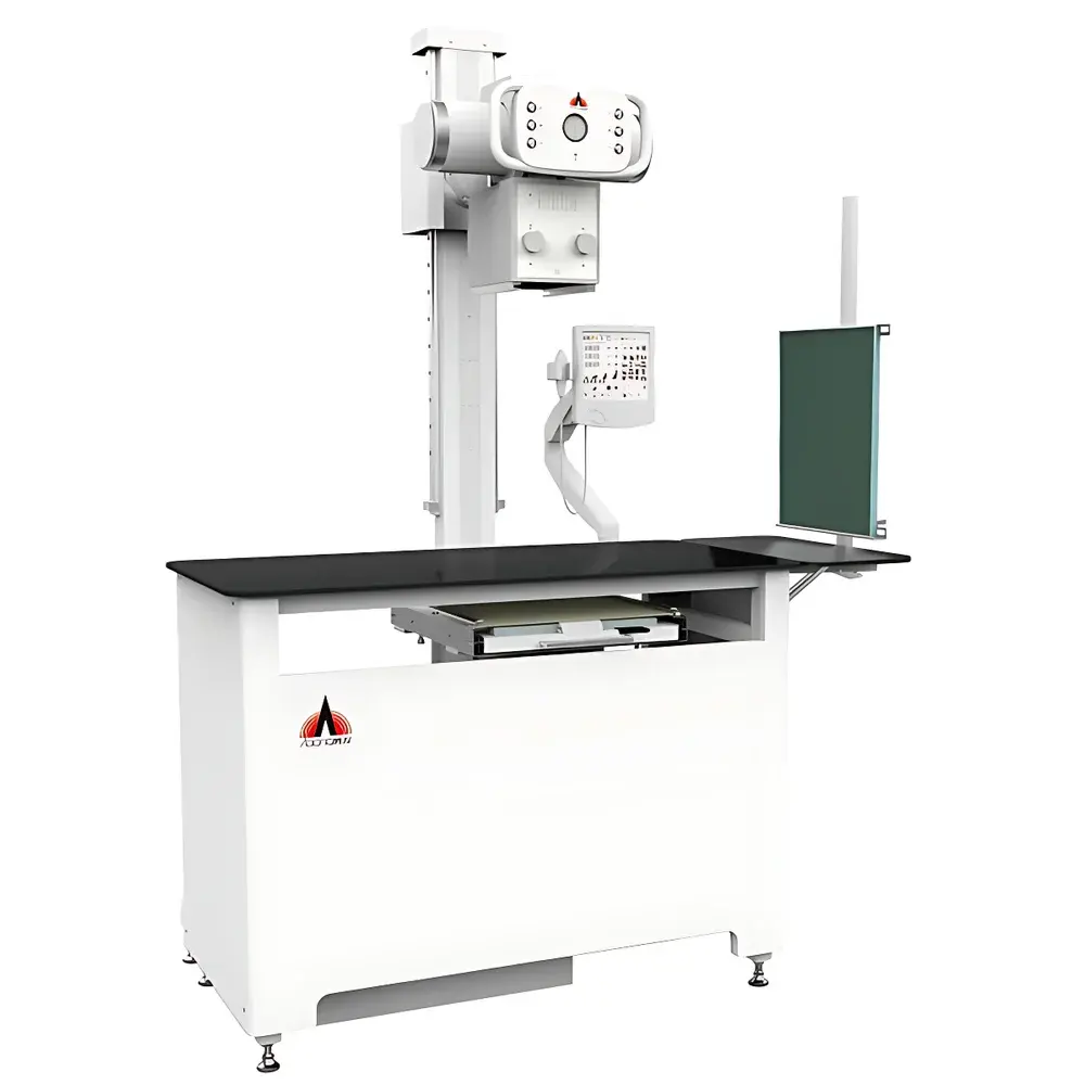
| Animal Table Dimensions | 120 × 60 × 90 cm (L×W×H) |
| Table Material | Carbon Fiber |
| Collimator Field Size (max) | 48 × 48 cm at SID = 100 cm |
| Light Field Duration | 60 s |
| Laser Pointer | Crosshair type |
| Collimator Adjustment | Manual knob |
| Grid Size | 17″ × 17″ (43 × 43 cm) |
| Grid Ratio | 8:1 |
| Grid Line Density | 103 lines/inch |
| Grid Focus Distance | 100 cm |
| Arm Vertical Travel | 74.5 cm |
| Arm Horizontal Travel | 50 cm |
| Tube Rotation Angle | ±90° |
Overview
The Aolong MXV100 Veterinary Digital Radiography System is a dedicated, floor-mounted X-ray imaging platform engineered for small- to medium-sized animal practices. It operates on the principle of projection radiography—utilizing a high-frequency, high-output X-ray generator and a precisely collimated beam to produce diagnostic-quality images with controlled dose delivery. Unlike conventional fixed-installation systems requiring structural reinforcement or trenching, the MXV100 employs a ground-clearance-free design with integrated mechanical stability, enabling immediate deployment in standard clinical rooms without architectural modification. Its human-centered ergonomics—including a 90 cm tabletop height and ±90° rotatable tube head—support consistent positioning across diverse species (canine, feline, avian, and exotic patients), while its carbon fiber table minimizes beam attenuation and secondary scatter, contributing to both image fidelity and ALARA-compliant radiation safety.
Key Features
- Integrated high-frequency X-ray generator (Delta Electronics): 32 kW output, 400 kHz operating frequency, optimized for compact veterinary environments; mounted beneath the table to preserve floor space.
- Rotatable X-ray tube assembly: ±90° angular adjustment enables oblique, lateral, and dorsoventral projections without repositioning the patient or equipment.
- Removable anti-scatter grid: 17″ × 17″ (43 × 43 cm), 8:1 ratio, 103 lines/inch, focused at 100 cm SID—designed to suppress Compton scatter and enhance contrast resolution in thick-tissue studies.
- Collimator with dual-function illumination: High-intensity light field (60 s duration) and crosshair laser alignment system ensure precise field localization and reproducible centering—critical for standardized orthogonal views in orthopedic and thoracic assessments.
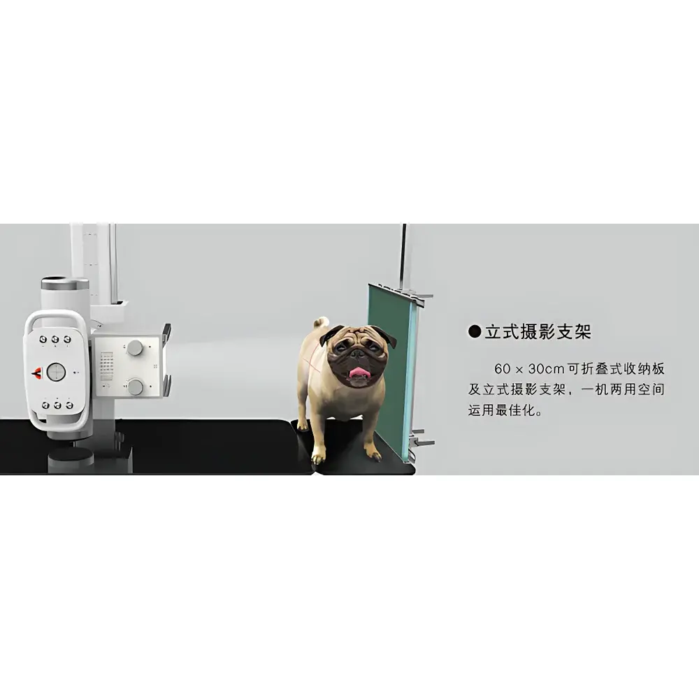
- Ergonomic, foldable upright Bucky stand: 60 × 30 cm carbon fiber detector tray accommodates DR panels up to 35 × 43 cm; supports standing, weight-bearing, and vertical projections without auxiliary support structures.
- Intuitive control panel with customizable positioning templates: Patented graphic overlays allow rapid visual reference for common anatomical projections (e.g., hip extended, stifle flexed, skull ventrodorsal), reducing setup time and inter-operator variability.
- Carbon fiber tabletop: Low atomic number composition ensures minimal X-ray absorption (<0.5% attenuation at 80 kV), preserving photon flux for optimal detector quantum efficiency and lowering required exposure parameters.
Sample Compatibility & Compliance
The MXV100 accommodates animals weighing up to 45 kg on its reinforced carbon fiber table, with adjustable restraint-compatible surface geometry. Its variable SID (source-to-image distance) capability—from 80 cm to 120 cm—and wide collimation range (up to 48 × 48 cm at 100 cm SID) permit adaptation to species-specific anatomy and detector formats (including wireless DR plates and tethered flat-panel detectors). The system complies with IEC 60601-1 (Medical Electrical Equipment Safety) and IEC 61331-1 (Protective Devices Against Ionizing Radiation); its grid focus distance and beam alignment meet ASTM E94 requirements for radiographic sensitivity. While not FDA-cleared as a Class II device (as applicable to U.S.-distributed veterinary systems), the MXV100’s design aligns with ISO 17025 traceability expectations for QA/QC in accredited imaging labs and supports GLP documentation workflows through configurable exposure logs.
Software & Data Management
The MXV100 interfaces seamlessly with DICOM-compliant PACS and third-party veterinary imaging software via Gigabit Ethernet. Exposure metadata—including kV, mA, mAs, focal spot size, grid usage, and collimation settings—is embedded in each DICOM header, ensuring auditability and consistency in longitudinal case review. Optional integration with RIS modules permits automatic exam protocol assignment based on species, body region, and clinical indication. All exposure parameters are stored locally with timestamp, operator ID, and detector serial number—supporting internal QA reviews and facilitating compliance with regional radiation safety reporting mandates (e.g., UK IR(ME)R, EU BSS Directive 2013/59/Euratom).
Applications
- Orthopedic evaluation: Hip dysplasia scoring (PennHIP, OFA), fracture characterization, patellar luxation grading, and post-operative hardware assessment.
- Thoracic imaging: Pulmonary pattern analysis, cardiac silhouette measurement (vertebral heart score), tracheal collapse staging, and mediastinal mass localization.
- Abdominal surveys: Gastrointestinal obstruction detection, urolith identification and localization, hepatic size estimation, and adrenal gland morphology screening.
- Dental radiography: Full-mouth series acquisition using angled tube positioning and customized collimation—minimizing overlap and maximizing root detail.
- Exotic and avian imaging: Low-dose, high-contrast protocols enabled by carbon fiber support and fine kV/mAs control—suitable for chelonians, psittacines, and lagomorphs.
FAQ
Is the MXV100 compatible with wireless DR detectors?
Yes—the system supports all major wireless DR detectors with standard DICOM SCP/SCU communication and includes configurable AET (Automatic Exposure Tracking) profiles per detector model.
Does the system include dose monitoring tools?
It records and exports per-exposure air kerma estimates (calculated from kV, mAs, filtration, and SID) in accordance with IEC 62494-1, enabling practice-level dose trend analysis.
Can the upright Bucky be used for weight-bearing studies?
Yes—the 60 × 30 cm carbon fiber plate supports static weight-bearing views of stifle, tarsus, and carpus with full mechanical locking at any height between 60–140 cm.
What regulatory documentation is provided for international shipment?
CE Declaration of Conformity (IEC 60601-1, IEC 61331-1), RoHS compliance certificate, and full technical file summary are supplied with each unit.
Is remote service access supported?
The embedded diagnostic interface allows secure, encrypted remote connection for firmware updates and calibration verification—subject to local network policy and GDPR/PIPL-compliant data handling agreements.


