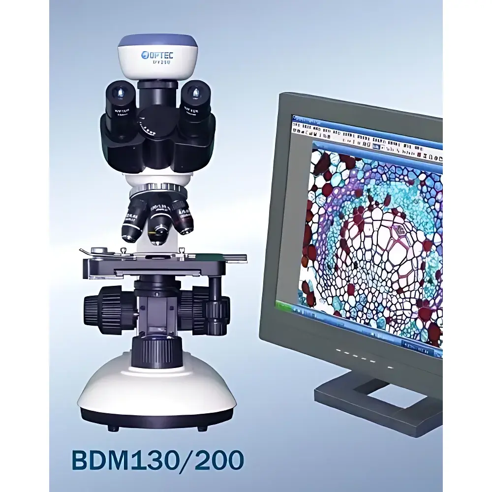
BDM300 Biological Microscope with Integrated Imaging Capability
| Origin | Beijing, China |
|---|---|
| Manufacturer Type | Authorized Distributor |
| Origin Category | Domestic (PRC) |
| Model | BDM300 |
| Price | USD 1,950 (FOB) |
| Magnification Range | 40×–1600× |
| Eyepieces | Wide-field Plan Eyepieces, 10×, Φ18 mm |
| Objective Lenses | Achromatic Plan Objectives (4×, 10×, 40× spring, 100× spring/oil) + Infinity-Corrected Achromatic Plan Objectives (4×, 10×, 20×, 40× spring, 100× spring/oil) |
| Observation Tube | Hinged Binocular Head (30° or 45° inclination, 360° rotation, interpupillary adjustment 50–75 mm) |
| Optional Trinocular Head (30° inclination, 1 | 5 beam splitter) |
| Nosepiece | Internal-Positioning Quadruple Revolving Nosepiece |
| Focus Mechanism | Coaxial Coarse/Fine Adjustment |
| Coarse Travel | 22 mm |
| Fine Adjustment | 0.2 mm/rev, Graduation: 2 µm |
| Stage | Mechanical Stage, 142 × 134 mm, X–Y Travel: 76 × 50 mm |
| Condenser | Abbe Condenser, NA 1.25, Adjustable Iris Diaphragm |
| Illumination | 6 V / 20 W Halogen Lamp (85–265 V AC input, dimmable) OR Optional LED Cold Light Source (rechargeable & non-rechargeable variants) |
| Accessories | Eyepieces (5×, WF16×, 10× measuring eyepiece [0.1 mm scale], 10× pointer eyepiece), Objectives (20×, 60× spring), Dual-Surface Mirror, Slide Micrometer (30 × 50 mm travel), C-Mount Adapters (0.5×, 1×), DSLR Camera Interfaces (NIKON, OLYMPUS, CANON), Digital Cameras (DV130: 1.3 MP, DV200: 2.0 MP), Darkfield Annuli (for 10× & 40× objectives) |
Overview
The BDM300 Biological Microscope is an upright, transmitted-light optical microscope engineered for high-fidelity observation and digital documentation of unstained and stained biological specimens in both fixed and live-cell contexts. Designed around an infinity-corrected optical pathway, the system supports modular expansion for phase contrast, darkfield, and basic fluorescence configurations—making it suitable for routine histology, cytology, microbiology, and introductory in vivo imaging workflows. Its dual-objective turret configuration accommodates both standard achromatic plan objectives and higher-resolution infinity-corrected optics, ensuring consistent image flatness and chromatic correction across the entire field of view. The instrument adheres to ISO 10934-1 (Microscopes — Nomenclature of components) and complies with IEC 61010-1 safety standards for laboratory electrical equipment. While not certified for GLP/GMP production environments, its mechanical stability, repeatable focus architecture, and traceable calibration pathways support QC/QA documentation requirements in academic, clinical training, and contract research laboratories.
Key Features
- Optical Architecture: Dual objective turret design supporting simultaneous use of finite and infinity-corrected achromatic plan objectives (4×–100×, oil immersion capable), minimizing chromatic aberration and field curvature.
- Precision Mechanics: Coaxial coarse/fine focusing with 2 µm fine-adjustment graduation, upper-limit stop, and adjustable tension control ensures reproducible Z-axis positioning during time-lapse or multi-plane imaging.
- Ergonomic Observation: Hinged binocular head with 30° (or optional 45°) inclination, 360° rotation, and 50–75 mm interpupillary adjustment reduces operator fatigue during extended sessions.
- Modular Illumination: Interchangeable 6 V / 20 W halogen source (broad-spectrum, CRI >95) or energy-efficient LED cold light system (CCT 5700 K, flicker-free PWM dimming) with dual power options (AC mains or internal battery).
- Digital Integration: Standard trinocular port with 1:5 beam-splitting ratio enables concurrent visual observation and high-fidelity image capture via C-mount–compatible cameras (DV130/DV200) or DSLR adapters for Nikon, Olympus, and Canon systems.
- Stage & Condenser: Large-area mechanical stage (76 × 50 mm travel) paired with NA 1.25 Abbe condenser and iris diaphragm ensures optimal Köhler illumination alignment and resolution control per magnification level.
Sample Compatibility & Compliance
The BDM300 accommodates standard 1 mm-thick glass microscope slides (76 × 26 mm) and petri dishes up to 100 mm diameter when used with optional stage inserts. It supports brightfield, darkfield (with included annuli for 10× and 40× objectives), and phase contrast (with add-on phase telescope and matching phase rings) modalities. Specimen thickness tolerance is ≤1.2 mm for cover-glass–mounted preparations. All optical components meet RoHS Directive 2011/65/EU material restrictions. Electrical compliance includes IEC 61010-1:2010 (Safety Requirements for Electrical Equipment for Measurement, Control, and Laboratory Use) and EMC conformity per IEC 61326-1:2020. The system does not carry CE marking for in vitro diagnostic (IVD) use or FDA 510(k) clearance; users deploying it in regulated clinical settings must validate performance per local SOPs and ISO/IEC 17025 requirements.
Software & Data Management
The BDM300 operates without proprietary firmware but interfaces seamlessly with industry-standard imaging software platforms including ImageJ/Fiji (open-source), NIS-Elements (Nikon), CellSens (Olympus), and LAS X (Leica). Digital camera outputs are delivered via USB 2.0 (DV130/DV200) or HDMI (optional upgrade), enabling direct acquisition into LabArchives ELN, Benchling, or institutional LIMS via metadata-tagged TIFF/RAW export. Audit-trail functionality—including timestamped image capture logs, objective ID auto-tagging, and exposure parameter recording—is achievable through third-party middleware compliant with FDA 21 CFR Part 11 when deployed with validated electronic signature modules. No embedded data storage or cloud synchronization is provided; all data persistence resides on user-controlled endpoints.
Applications
- Routine histopathological screening of H&E- and IHC-stained tissue sections.
- Live-cell monitoring of adherent mammalian cultures (e.g., HeLa, NIH/3T3) using low-magnification time-lapse sequences.
- Morphological identification of bacterial colonies, yeast, and protozoa in clinical microbiology education labs.
- Basic hematology analysis (e.g., peripheral blood smear differential counts) under standardized brightfield conditions.
- Plant anatomy studies, including stomatal density quantification and vascular bundle visualization in leaf cross-sections.
- Training platform for optical alignment procedures (Köhler illumination, centering condenser, adjusting critical aperture) per ASTM E2869-22 guidelines.
FAQ
Is the BDM300 compatible with fluorescence microscopy?
The base configuration does not include excitation/emission filters or a fluorescence illuminator. However, the infinity-corrected optical path and trinocular port allow integration of third-party epi-fluorescence attachments (e.g., Thorlabs LED-based modules) with appropriate filter cubes.
Can the microscope be used for oil immersion at 100× without damaging the lens?
Yes—the 100× objective features spring-loaded retraction and is designed for immersion oil (n = 1.518). Always clean the front lens immediately after use with lens-grade xylene and lint-free wipes to prevent residue buildup.
What is the maximum recommended camera sensor size for the 1× C-mount adapter?
The 1× adapter supports sensors up to 1/1.8″ format (8.9 × 6.7 mm). Larger sensors (e.g., APS-C) require the 0.5× reducer to avoid vignetting and maintain full-field illumination.
Does the LED illumination option support DIC or polarization?
No—neither the halogen nor LED source includes polarizers or Nomarski prisms. Polarization contrast requires separate analyzer and polarizer kits mounted in the filter slots above/below the condenser.
Is technical documentation available in English with metrological traceability statements?
Yes—calibration certificates for stage micrometers and eyepiece reticles are supplied with NIST-traceable reference standards. Full English-language operation manual, optical schematics, and ISO-compliance summary are provided upon request.


