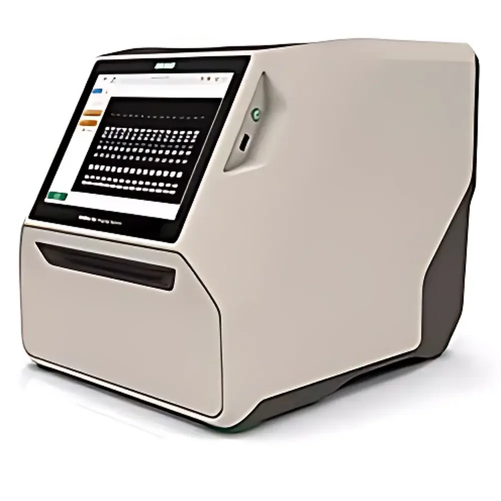
Bio-Rad GELDoc Go Gel Imaging System
| Origin | USA |
|---|---|
| Manufacturer Type | Authorized Distributor |
| Origin Category | Imported |
| Model | GELDoc Go |
| Instrument Type | Standard Gel Imaging System |
| CCD/CMOS Resolution | 1,024 × 768 pixels (touch display) / 6.3 MP scientific CMOS sensor |
| Bit Depth | 16-bit (65,536 gray levels) |
| Dynamic Range | >3.5 OD (optical density) |
| Sensor Size | 1/2" format |
| Detection Sensitivity | 0.02 mg protein (Coomassie-stained SDS-PAGE) |
| Signal-to-Noise Ratio | 56 dB |
| Lens | Motorized 8–48 mm zoom lens |
| Excitation Sources | Transmitted UV (254/302 nm, standard), Side white light (standard), Transmitted white light (optional, with white-light tray), Transmitted blue light (optional, with blue-light tray) |
| Emission Filter | 535–645 nm bandpass (standard) |
| Image Formats | 16-bit SCN, TIFF, JPEG |
| Touchscreen | 9.7" multi-touch LCD (1,024 × 768) |
| Imaging Area | 21 × 14 cm (W × H) |
| Dimensions (D × W × H) | 44.8 × 36.0 × 35.3 cm |
| Weight | ~16 kg |
| Operating Temperature | 10–28°C |
| Operating Humidity | 20–80% RH (non-condensing) |
| Power Supply | 100–240 VAC, 50–60 Hz |
Overview
The Bio-Rad GELDoc Go Gel Imaging System is a compact, integrated benchtop platform engineered for rapid, reproducible visualization and quantitative analysis of nucleic acid and protein electrophoresis gels, as well as Stain-Free gels and western blots. Built upon a high-sensitivity 6.3 megapixel scientific CMOS detector and motorized 8–48 mm zoom optics, the system captures images with 16-bit depth (65,536 gray levels) and a dynamic range exceeding 3.5 optical density (OD) units—enabling accurate quantification across broad signal intensities without saturation or loss of low-abundance band detail. Unlike legacy CCD-based systems, the GELDoc Go employs a modern CMOS architecture optimized for low-noise performance at ambient laboratory temperatures, delivering consistent sensitivity down to 0.02 mg of Coomassie-stained protein per band. Its optical path integrates dual excitation modalities—transmitted UV (254/302 nm) and side-mounted white light—as standard, with optional transmitted white and blue light modules supporting diverse fluorescent and chemiluminescent detection strategies. The system operates on a self-contained architecture with no external PC dependency for image acquisition, making it ideal for shared core facilities, teaching labs, and QC environments where footprint, ease of use, and regulatory compliance are critical.
Key Features
- Integrated 9.7-inch multi-touch display (1,024 × 768 resolution) enabling fully standalone operation—no external computer required for image capture or basic annotation.
- Smart tray recognition technology automatically detects tray type (UV, Stain-Free, white-light, or blue-light) and configures optimal illumination, exposure, and focus parameters without user input.
- Pre-calibrated motorized auto-focus across full zoom range ensures consistent sharpness from wide-field gel overview to high-magnification band inspection.
- Dual auto-exposure algorithms—“Optimized” (for maximum dynamic range and quantitative fidelity) and “Quick” (for rapid preview and documentation)—adapt to sample type and staining method in real time.
- Modular excitation design supports regulatory-compliant workflows: UV transillumination meets ASTM E2559 for nucleic acid visualization; Stain-Free imaging complies with USP guidelines for protein quantification without dye interference.
- Robust mechanical housing (16 kg net weight) and thermally stable optical train ensure measurement repeatability across shifts and environmental fluctuations typical in ISO 17025-accredited laboratories.
Sample Compatibility & Compliance
The GELDoc Go accommodates standard mini-gels (up to 14 × 21 cm), including polyacrylamide (PAGE), agarose, and gradient gels, as well as nitrocellulose and PVDF membranes for western blotting. It natively supports ethidium bromide, SYBR Safe, GelRed, Coomassie Blue, Silver Stain, and Bio-Rad’s proprietary Stain-Free technology. For regulated environments, the system aligns with key quality frameworks: Image Lab Touch software implements role-based user authentication, electronic signatures, and full audit trails compliant with FDA 21 CFR Part 11, EU Annex 11, and GLP/GMP data integrity requirements. All image metadata—including exposure time, lens position, filter selection, and calibration timestamp—is embedded in the 16-bit SCN file format and preserved through export to TIFF or CSV. No image processing occurs prior to storage; raw acquisition data remains immutable and traceable throughout the analytical lifecycle.
Software & Data Management
Two complementary software environments are provided: Image Lab Touch (embedded on-device) and Image Lab PC (Windows-based). Image Lab Touch delivers intuitive one-tap acquisition, real-time contrast/brightness adjustment, region-of-interest (ROI) drawing, molecular weight estimation, and lane/band profiling directly on the touchscreen. Image Lab PC extends functionality with advanced densitometry, multiplex normalization (e.g., loading control correction), statistical comparison across multiple gels, and direct export to Excel-compatible .csv or structured database formats (e.g., SQL via ODBC). Both versions enforce strict data governance: all user actions—including parameter changes, annotations, and export events—are logged with timestamps and operator IDs. Software validation packages, IQ/OQ documentation templates, and electronic record retention protocols are available to support laboratory accreditation under CLIA, CAP, or ISO/IEC 17025.
Applications
- Quantitative analysis of DNA fragments in agarose and polyacrylamide gels for genotyping, PCR product verification, and restriction digest validation.
- Densitometric quantification of protein expression levels in SDS-PAGE and western blots, including post-translational modification assessment via band shift analysis.
- Stain-Free total protein normalization for western blotting—eliminating antibody-based housekeeping controls and reducing inter-assay variability.
- Documentation and archiving of electrophoretic results in GLP-compliant toxicology studies and biopharmaceutical process development reports.
- Teaching laboratory instruction in molecular biology techniques, where rapid setup, visual feedback, and standardized output facilitate student learning and grading consistency.
FAQ
Does the GELDoc Go require a dedicated computer for image acquisition?
No. All image capture, preview, annotation, and export functions operate natively on the integrated touchscreen. A PC is only needed for advanced analysis using Image Lab PC software.
Can the system be validated for use in FDA-regulated environments?
Yes. With Image Lab Secure software (optional upgrade), the system supports 21 CFR Part 11 compliance, including electronic signatures, audit trail review, and secure user access controls.
What is the minimum detectable protein mass under Coomassie staining?
The system achieves reliable detection of ≥0.02 mg protein per band on standard SDS-PAGE gels, verified per Bio-Rad Application Note 10022195.
Is calibration required before each use?
No. The system performs automatic optical calibration during startup. Optional annual hardware calibration services are available through Bio-Rad Field Service Engineers.
Are third-party filters or trays supported?
Only Bio-Rad-certified imaging trays and emission filters are validated for performance and regulatory compliance. Use of non-OEM components voids warranty and may compromise data integrity.


