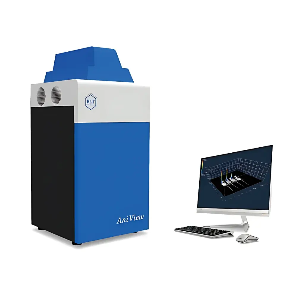
BioLight PlantView100 Plant In Vivo Imaging System
| Brand | BioLight |
|---|---|
| Origin | Guangdong, China |
| Model | PlantView100 |
| Imaging Modality | Optical Bioluminescence & Fluorescence Imaging |
| Maximum Field of View | 280 mm × 280 mm |
| Camera | Dual-cooled scientific-grade CCD (−100 °C), QE > 95% |
| Excitation Sources | Up to 20 narrowband high-power LEDs |
| Emission Filters | Up to 10 precision OD6 bandpass filters |
| Optional Modules | Side-view imaging module, X-ray imaging module, Chlorophyll fluorescence module, Light simulation module (4-channel LED: blue/white/red/NIR), Cell labeling quantification module (tube & 96-well formats) |
| Software | GLP-compliant, audit-trail enabled, auto-calibrated photon flux quantification (p/s/cm²/sr), raw + processed data segregation, export to CSV/Excel with embedded metadata |
Overview
The BioLight PlantView100 Plant In Vivo Imaging System is a dedicated optical bioluminescence and fluorescence imaging platform engineered for non-invasive, longitudinal monitoring of biological processes in intact, living plants. It operates on the principle of photon detection from luciferase-based bioluminescent reporters (e.g., Firefly Luc, Renilla Luc) and fluorescent protein tags (e.g., GFP, mCherry, YFP) or exogenous dyes, enabling spatially resolved, quantitative measurement of gene expression dynamics, protein–protein interactions, pathogen colonization, stress responses, and circadian rhythms at the whole-plant level. Unlike conventional microscopy or destructive sampling methods, the PlantView100 captures signal emission across the entire plant architecture—including roots, stems, leaves, flowers, and seeds—without tissue disruption, thereby preserving physiological context and temporal continuity. Its deep-cooled, high-quantum-efficiency CCD sensor and ultra-low-noise optical path support detection of sub-picowatt-level photon fluxes, making it suitable for low-expression transgenic lines and early-stage infection or developmental events. The system integrates tightly controlled environmental simulation (light spectrum, intensity, timing) and modular hardware expansion, positioning it as a core infrastructure tool for modern plant systems biology, functional genomics, and translational breeding programs.
Key Features
- Ultra-large field-of-view imaging: Dual-camera configuration supports simultaneous top-view (280 mm × 280 mm) and optional side-view acquisition, enabling full-plant imaging of Arabidopsis, rice, maize seedlings, and multi-sample trays (e.g., 96-well plates, Petri dishes, germination boxes).
- High-sensitivity photon detection: Scientific-grade back-illuminated CCD cooled to −100 °C; quantum efficiency >95% at 600–700 nm; integrated OD6 emission filters and background subtraction algorithms ensure high signal-to-noise ratio even under ultra-low-light conditions.
- Multi-spectral excitation flexibility: 20 independently controllable narrowband LED sources (365–740 nm), each with stable output and minimal spectral drift; paired with 10 precision bandpass emission filters for multiplexed fluorescence unmixing and autofluorescence rejection.
- Programmable light simulation module: Dual LED panels delivering calibrated blue (450 nm), white (5000 K), red (660 nm), and near-infrared (730 nm) irradiance; software-controlled intensity (0–100%) and photoperiod scheduling enable precise replication of photomorphogenic and circadian experimental regimes.
- Modular expandability: Optional add-ons include side-view imaging station (with motorized rotation stage and high-throughput sample adapter), X-ray transmission module (for internal structural and hydration mapping), chlorophyll fluorescence imaging (Fv/Fm, ΦPSII), and cell-labeling quantification unit (tube- and microplate-based luminometric calibration curves).
Sample Compatibility & Compliance
The PlantView100 accommodates diverse botanical specimens—from single seeds and callus cultures to mature rosettes and hydroponically grown root systems—within its standardized dark chamber. Sample holders are compatible with standard labware (e.g., 100 mm Petri dishes, 96-well plates, custom acrylic trays). All optical and mechanical components comply with IEC 61000-6-3 (EMC immunity) and IEC 61000-6-4 (EMC emissions) standards. The system’s software architecture conforms to Good Laboratory Practice (GLP) requirements per OECD Series 125 and ISO/IEC 17025:2017 Annex A, including mandatory audit trails, electronic signatures (21 CFR Part 11-ready configuration), immutable raw data archiving, and separation of original acquisition files from processed datasets. Quantitative outputs are traceable to SI-derived radiometric units (photons per second per square centimeter per steradian, p/s/cm²/sr), ensuring cross-platform reproducibility and regulatory acceptability in academic, industrial, and contract research settings.
Software & Data Management
BioLight ImageStudio Plant Edition is a purpose-built, Windows-native application offering fully automated instrument control, protocol-driven acquisition, and compliant data handling. Users define exposure time, binning, lens aperture, LED intensity, filter position, and stage height via intuitive graphical workflows. Preconfigured templates cover common assays (e.g., “Luciferase Time-Course”, “GFP Expression Screening”, “Chlorophyll Fv/Fm Mapping”). All operations generate timestamped metadata logs, including hardware state, ambient temperature, and user identity. Image quantification employs region-of-interest (ROI) masking, background correction using rolling-ball or polynomial fitting, and spectral unmixing based on reference spectra. Results are exported as Excel-compatible CSV files containing absolute photon flux values, statistical summaries (mean ± SD), and embedded thumbnails. Raw TIFFs retain EXIF-style headers with acquisition parameters, satisfying FAIR (Findable, Accessible, Interoperable, Reusable) data principles. Software updates are provided free of charge for the instrument’s lifetime, with version-controlled release notes and validation reports available upon request.
Applications
- Transgenic line screening: High-throughput identification of luciferase- or fluorescent-protein-expressing plants during early seedling stages, reducing false negatives and accelerating T0–T2 selection cycles.
- Circadian and photoperiodic studies: Real-time tracking of clock gene promoters (e.g., CCA1::LUC, TOC1::LUC) under programmable light/dark regimes to dissect entrainment kinetics and phase shifts.
- Plant–pathogen interaction analysis: Spatial mapping of bioluminescent bacterial or fungal colonization (e.g., Pseudomonas syringae, Magnaporthe oryzae) and host defense activation (e.g., PR1::LUC, NPR1::GFP) over 72+ hours.
- Abiotic stress phenotyping: Quantitative assessment of ROS-sensitive reporters (e.g., HyPer), antioxidant enzyme promoters (e.g., APX2::LUC), or osmotic stress markers (e.g., RD29A::LUC) following drought, salinity, or heavy metal exposure.
- Root architecture dynamics: Side-view imaging combined with time-lapse reconstruction enables morphometric analysis of lateral root emergence, gravitropism, and hydrotropism without soil disturbance.
- Seed germination and vigor assays: Multiplexed luminescence readouts from embryo-specific promoters (e.g., ABI3::LUC) correlate with metabolic reactivation and stress resilience prior to radicle emergence.
FAQ
What is the minimum detectable photon flux for bioluminescent imaging?
The system achieves a theoretical detection limit of ≤10⁴ photons/s/cm²/sr under optimal conditions (10-minute exposure, −100 °C sensor, OD6 filter, no background light leakage). Actual sensitivity depends on reporter brightness, tissue depth, and chlorophyll absorption.
Can the PlantView100 perform kinetic imaging over multiple days?
Yes. Automated stage positioning, scheduled LED activation, and thermal-stabilized chamber design allow unattended sequential imaging at user-defined intervals (minutes to 24-hour periods) for up to 14 days.
Is the software validated for regulated environments (e.g., GLP, GMP)?
The software includes configurable 21 CFR Part 11 compliance features (electronic signatures, audit trail, role-based access control) and meets OECD GLP data integrity requirements. Full validation documentation is available under NDA.
Does the system support spectral unmixing for multiple fluorescent proteins?
Yes. With ≥3 excitation/emission channel combinations and built-in linear unmixing algorithms, users can resolve overlapping signals from GFP/mCherry/CyanFP simultaneously when appropriate reference spectra are provided.
How is temperature controlled during long-term imaging?
The chamber incorporates active Peltier cooling and optional water-circulation interface (via external chiller) to maintain ambient stability within ±0.5 °C during extended acquisitions, critical for circadian and developmental studies.
Related Products


