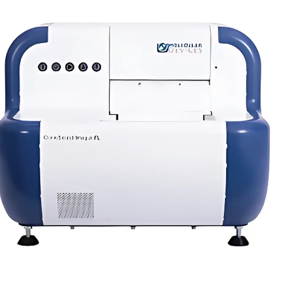
CloneSelect Imager FL Fluorescence and Brightfield Cell Growth Imaging System
| Brand | Molecular Devices |
|---|---|
| Origin | Shanghai, China |
| Manufacturer Type | Authorized Distributor |
| Origin Category | Domestic (China-manufactured) |
| Model | CloneSelect Imager FL |
| Pricing | Available Upon Request |
Overview
The CloneSelect Imager FL is a high-throughput, non-invasive cell growth analysis system engineered for precision monitoring of monoclonal colony formation and proliferation kinetics in microplate formats. Leveraging simultaneous multi-channel fluorescence and brightfield imaging, it enables label-free, longitudinal assessment of cell confluence, morphology, and reporter expression (e.g., GFP, RFP) without enzymatic assays or cytotoxic dyes. Designed specifically for biopharmaceutical development workflows, the system supports critical quality attributes required under ICH Q5D and regulatory expectations for clonality assurance—particularly during master cell bank (MCB) generation and IND-enabling studies per 21 CFR Part 312. Its optical architecture integrates high-sensitivity CMOS sensors, motorized filter wheels, and automated focus calibration to ensure consistent image acquisition across 6–384-well plates, accommodating both adherent (e.g., CHO-K1, HEK293) and suspension-adapted cell types (e.g., hybridomas, iPSCs).
Key Features
- Simultaneous dual-mode imaging: High-resolution brightfield + up to four independent fluorescence channels (ex/em configurable via interchangeable filter sets)
- Automated clonality verification: “Loci of Growth” algorithm identifies single-cell-derived colonies at Day 0 and tracks lineage expansion over time
- Quantitative confluence and cell count analytics: Pixel-based segmentation delivers reproducible metrics for each well, including % confluence, estimated cell number, and growth rate (doubling time)
- FDA-submission-ready reporting: Structured, audit-trail-enabled Monoclonality Report generation compliant with ALCOA+ principles and 21 CFR Part 11 data integrity requirements
- Plate-level metadata integration: Full traceability of imaging parameters (exposure time, gain, focus offset), environmental conditions (if integrated with incubator), and user actions
- Flexible workflow compatibility: Supports manual or automated plate loading; compatible with standard SBS-format microplates and stackers
Sample Compatibility & Compliance
The CloneSelect Imager FL accommodates a broad spectrum of mammalian cell systems used in therapeutic protein production and cell therapy development—including CHO, HEK293, hybridoma, CAP-T, and pluripotent stem cell lines—regardless of adherence phenotype. It meets essential performance criteria outlined in ASTM E3157-18 (Standard Guide for Evaluation of Automated Cell Culture Systems) and aligns with ISO/IEC 17025 requirements for measurement uncertainty in image-based quantitation. All software-generated reports include embedded timestamps, electronic signatures, and immutable audit logs required for GLP/GMP-regulated environments. The system supports IQ/OQ documentation packages and facilitates validation against USP and ICH Q5B guidelines for clonal derivation evidence.
Software & Data Management
Powered by CloneSelect Software v5.x, the platform provides a secure, role-based interface for acquisition, analysis, and report export. Raw images are stored in vendor-neutral TIFF format with embedded EXIF metadata; processed data (confluence curves, cell counts, growth rates) are saved in HDF5 containers supporting programmatic access via Python APIs. The software enforces 21 CFR Part 11 compliance through electronic signature workflows, automatic session logging, and protected audit trails that record all parameter modifications, image reprocessing events, and report generation actions. Export options include PDF/A-2b (for long-term archival), Word (.docx), and CSV for downstream statistical analysis in JMP or GraphPad Prism.
Applications
- Monoclonal antibody development: Objective confirmation of single-cell origin for MCB establishment and regulatory filings
- CRISPR/Cas9 and other gene-editing validation: Multiplexed fluorescent reporter tracking to confirm on-target editing and isolate edited clones
- Cell line development (CLD): Optimization of seeding density, media composition, and clone selection criteria based on kinetic growth profiling
- Toxicity and potency assays: Label-free confluence-based IC50 determination in response to small molecules, biologics, or viral vectors
- iPSC differentiation monitoring: Quantitative assessment of morphological transitions and marker expression dynamics across multiwell arrays
- Process characterization studies: Correlation of early-passage growth behavior with late-stage productivity and stability metrics
FAQ
Does the CloneSelect Imager FL require fluorescent labeling or cell staining?
No—its brightfield and autofluorescence-capable fluorescence channels enable label-free confluence measurement and endogenous reporter detection (e.g., GFP/RFP), eliminating reliance on viability dyes or enzymatic substrates.
Can the system validate clonality for FDA submission under 21 CFR Part 312?
Yes—the Monoclonality Report module generates structured, timestamped, electronically signed documentation meeting FDA expectations for IND-enabling clonality evidence, including Day 0 single-cell imaging and lineage tracking.
Is the software compliant with 21 CFR Part 11 for electronic records and signatures?
Yes—audit trails, electronic signatures, and system access controls are fully implemented per Part 11 Annex A guidance, supporting inspection-readiness in regulated biomanufacturing environments.
What microplate formats does it support?
Standard SBS-compliant plates from 6- to 384-well, including opaque, clear-bottom, and black-walled configurations for fluorescence applications.
How is data integrity ensured during long-term imaging experiments?
All acquisitions are checksum-verified upon save; raw images retain original sensor metadata; processed datasets are version-controlled and linked to source images via UUID-based referencing.


