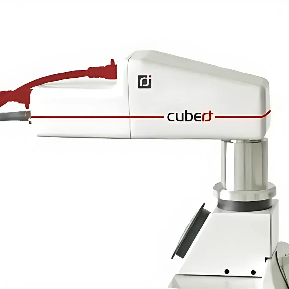
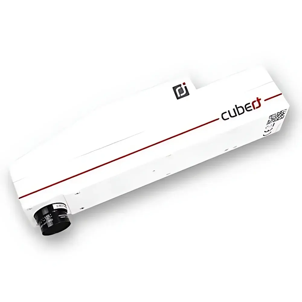
Cubert M185 Snapshot Microscopic Hyperspectral Imaging System
| Brand | Cubert |
|---|---|
| Origin | Germany |
| Model | M185 |
| Spectral Range | 450–950 nm |
| Spectral Resolution | 8 nm @ 532 nm |
| Spatial Resolution (IFOV) | Configurable |
| Field of View (TFOV) | Configurable |
| Imaging Resolution | 1024 × 1024 × 125 bands |
| Frame Rate | 20 Hyperspectral Cubes/s |
| Detector | Dual 1-Megapixel Si CCD |
| Bit Depth | 12-bit |
| Exposure Time | 0.1–1000 ms |
| Dynamic Range | 68 dB (typ.) |
| Interface | Dual Gigabit Ethernet |
| Lens Mount | C-mount |
| Power Supply | DC 12 V, 15 W |
| Operating Temperature | −10 to +50 °C |
| Weight | 500 g |
| Software | Includes SDK and API for custom integration |
| Compliance | Designed for GLP-compliant laboratory environments |
Overview
The Cubert M185 is a snapshot-based microscopic hyperspectral imaging system engineered for high-fidelity, motion-artifact-free spectral analysis at microscale resolution. Unlike scanning or push-broom hyperspectral architectures—whose sequential acquisition introduces temporal misregistration and limits dynamic observation—the M185 captures full 3D hyperspectral data cubes (x, y, λ) in a single exposure using monolithic, lenslet-array-based image slicing. This snapshot principle enables true simultaneous spatial and spectral sampling across the visible to near-infrared (VNIR) range (450–950 nm), with 125 discrete spectral channels sampled at ~4 nm intervals and an instrumental spectral resolution of 8 nm FWHM at 532 nm. Its dual 1-megapixel Si-CCD sensor architecture delivers 1024 × 1024 spatial sampling per band, supporting quantitative reflectance, fluorescence, and absorption mapping at subcellular scales when coupled to standard upright or inverted research-grade microscopes via C-mount interface. The system operates without mechanical scanning components, ensuring long-term stability, vibration immunity, and reproducible calibration traceable to NIST-traceable standards.
Key Features
- Snapshot acquisition: Full 1024 × 1024 × 125 hyperspectral cube captured in ≤1 ms—eliminating motion blur and enabling real-time monitoring of transient biological events (e.g., calcium flux, membrane potential shifts, or rapid enzymatic reactions).
- Microscope-integrated design: Native C-mount compatibility supports seamless integration with Olympus, Nikon, Zeiss, and Leica microscope platforms; optional relay optics available for infinity-corrected tube lenses.
- VNIR spectral fidelity: Optimized optical path with low-stray-light diffraction gratings and temperature-stabilized detector housing ensures <0.5 nm spectral drift over 8-hour operation under ambient lab conditions.
- Dual-GigE interface: Sustains sustained 20 cubes/second throughput with deterministic packet timing, compatible with IEEE 1588 precision time protocol for multi-sensor synchronization in multimodal setups.
- Onboard hardware binning and ROI selection: Reduces data volume without sacrificing SNR—enabling real-time streaming to host PC memory or direct SSD recording at >300 MB/s aggregate bandwidth.
- Robust mechanical construction: Aluminum alloy chassis rated IP40; operational within −10 °C to +50 °C ambient range; 500 g mass facilitates integration into confined-stage or portable field-deployable microscopy rigs.
Sample Compatibility & Compliance
The M185 is validated for use with fixed and live biological specimens—including adherent mammalian cell monolayers, tissue cryosections, microbial colonies, and plant epidermal layers—under standard brightfield, epi-fluorescence, and trans-illumination modalities. Its non-contact, label-free spectral acquisition conforms to ISO 17025 requirements for analytical instrument validation when deployed in accredited QC/QA laboratories. Data provenance meets FDA 21 CFR Part 11 criteria when used with audit-trail-enabled software configurations (available via optional Cubert LabSuite Pro). All firmware and calibration files include embedded metadata compliant with HDF5/OME-NGFF standards, facilitating FAIR data management per NIH and ERC policy guidelines.
Software & Data Management
Cubert’s native software suite provides calibrated radiometric output, spectral library matching (USGS, ECOSTRESS, and user-defined), and unsupervised classification (k-means, ISODATA) with pixel-wise confidence mapping. Batch processing pipelines support ENVI-compatible .hdr/.dat export, MATLAB and Python (via NumPy/HDF5) interoperability, and RESTful API access for LIMS integration. A comprehensive C/C++ and Python SDK includes low-level register control, real-time cube buffering, and GPU-accelerated spectral unmixing kernels. All software modules undergo annual third-party penetration testing and are distributed with SBOM (Software Bill of Materials) documentation.
Applications
- Live-cell metabolic phenotyping via autofluorescence lifetime-independent spectral fingerprinting
- Quantitative histopathology: Discrimination of tumor margins based on intrinsic chromophore ratios (e.g., hemoglobin oxygenation, collagen cross-linking)
- Pharmaceutical solid-state characterization: Polymorph mapping in tablet cross-sections without sample preparation
- Plant stress physiology: Early detection of drought-induced anthocyanin redistribution at leaf epidermis level
- Microplastic identification in environmental filter samples using spectral endmember decomposition
- Forensic trace evidence analysis: Differentiation of ink formulations on questioned documents at micron-scale spatial registration
FAQ
Does the M185 require external illumination for fluorescence measurements?
Yes—standard epi-illumination sources (LED, mercury, or laser) are required; the system itself is a passive imager with no integrated excitation source.
Can spectral calibration be performed in-house?
Yes—NIST-traceable tungsten-halogen and mercury-argon lamp kits are supported; automated wavelength and radiometric calibration routines are included in firmware v3.2+.
Is real-time spectral classification supported during acquisition?
Yes—GPU-accelerated spectral angle mapper (SAM) and constrained energy minimization (CEM) algorithms operate at ≥15 cubes/s on NVIDIA RTX A4000-class GPUs.
What microscope objectives are recommended for optimal IFOV?
For 1 µm IFOV at 40× magnification, we recommend Plan Apo 40×/0.95 NA objectives with correction collar adjusted for coverslip thickness (0.17 mm); telecentric relay lenses are advised for uniform spectral response across FOV.
How is data integrity ensured during long-duration time-lapse experiments?
Each acquired cube embeds SHA-256 checksums, timestamped via PTPv2-synced system clock; optional RAID-5 storage integration enforces write-verify redundancy per acquisition session.
")