Delmic Meteor 2.0 Integrated Cryo-Fluorescence Light Microscope for Correlative Cryo-FIB/SEM
| Brand | Delmic |
|---|---|
| Origin | Netherlands |
| Model | Meteor / Meteor 2.0 |
| Integration Type | In-chamber cryo-fluorescence light microscope (cryo-FLM) for direct mounting on cryo-FIB/SEM dual-beam systems |
| Vacuum Compatibility | High-vacuum compatible (≤1×10⁻⁶ mbar) |
| Optical Resolution | Subcellular (≤300 nm lateral, dependent on NA and wavelength) |
| Field of View | Up to 500 µm × 400 µm (tile-scanned, stitched) |
| Imaging Modes | Widefield fluorescence, Z-stack acquisition, multi-channel sequential imaging |
| Excitation Sources | LED-based, configurable wavelengths (e.g., 405 nm, 488 nm, 561 nm, 640 nm) |
| Emission Filters | Motorized filter wheel with bandpass filters (FWHM ≤25 nm), minimal spectral crosstalk |
| Objective | High-NA apochromatic cryo-objective (e.g., 0.95 NA, 20× or 40×, corrected for ice thickness) |
| Detector | Back-illuminated sCMOS camera (QE >95% at 550 nm, read noise <1.5 e⁻ rms) |
| Software Platform | ODEMIS (open-source, Python-based), including ODEMIS Advanced for automated correlative workflows |
| Compliance | Designed for GLP/GMP-aligned cryo-EM workflows |
Overview
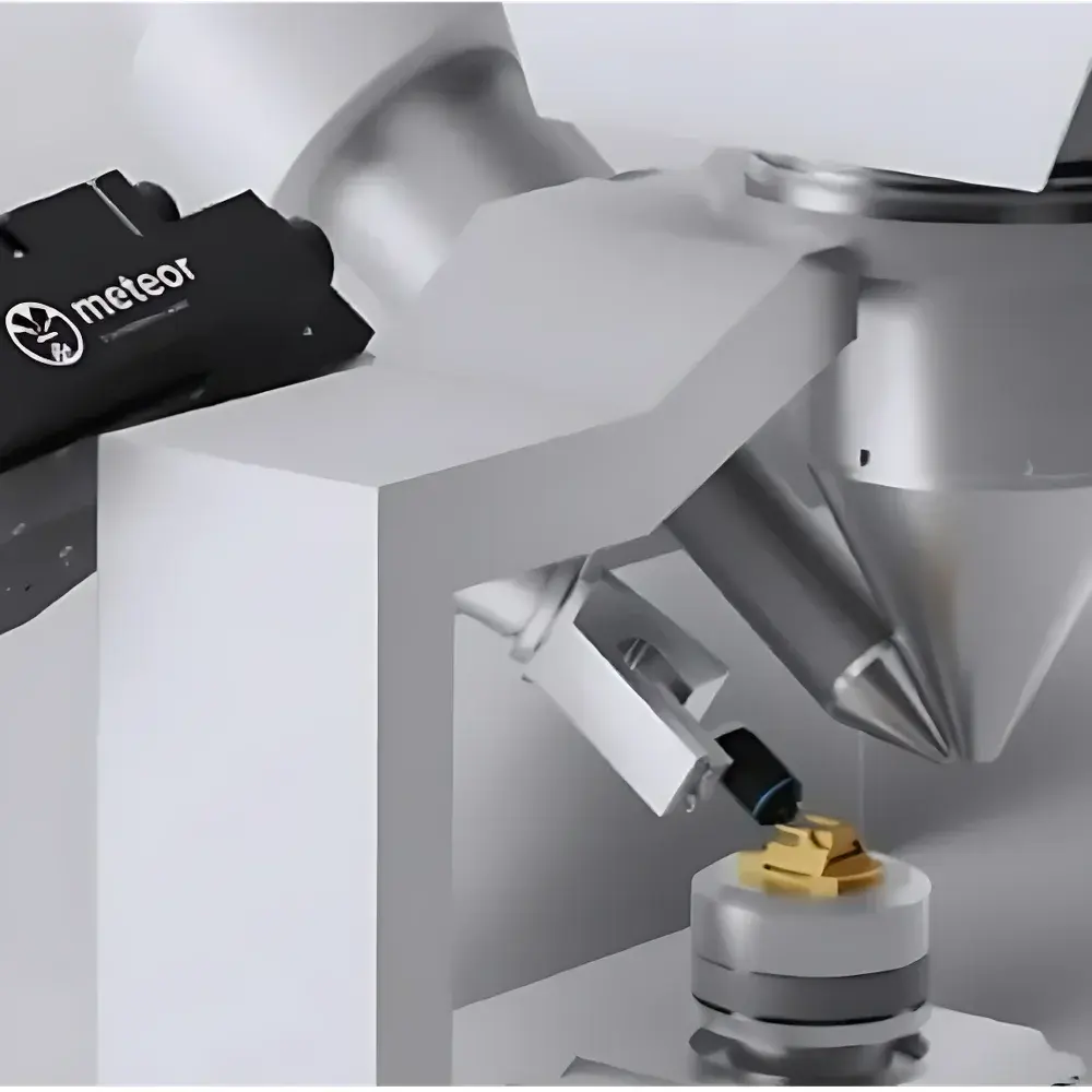
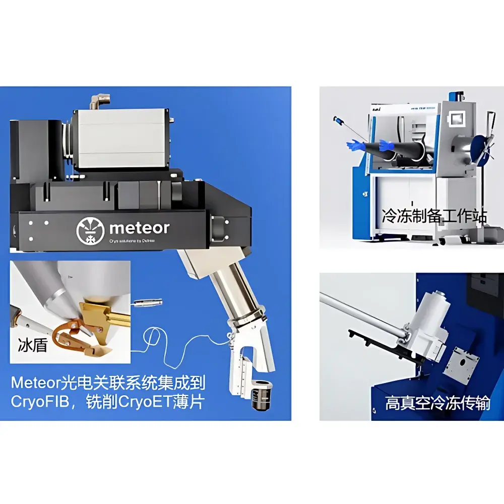
The Delmic Meteor 2.0 is an in-chamber cryo-fluorescence light microscope (cryo-FLM) engineered for direct integration into high-vacuum cryo-focused ion beam/scanning electron microscope (cryo-FIB/SEM) dual-beam systems. It enables true in situ cryo-correlative light and electron microscopy (cryo-CLEM), eliminating the need for intermediate sample transfers between standalone cryo-FLM and cryo-FIB instruments. By performing fluorescence localization directly inside the FIB/SEM chamber—under stable cryogenic conditions (<−170 °C) and high vacuum (≤1×10⁻⁶ mbar)—the Meteor 2.0 mitigates two principal failure modes in cryo-electron tomography (cryo-ET) workflows: ice contamination and ROI misregistration. Its optical architecture leverages high-numerical-aperture (NA ≥0.95) apochromatic objectives corrected for ice-layer dispersion, coupled with back-illuminated sCMOS detection and low-noise LED excitation, enabling robust photon collection from vitrified biological specimens—even after FIB milling, where residual fluorescence signals may be attenuated by up to two orders of magnitude.
Key Features
- In-chamber fluorescence imaging: Fully integrated optical path mounted on a dedicated port of the cryo-FIB/SEM, operating under high vacuum without compromising chamber integrity or cooling stability.
- Subcellular-resolution widefield imaging: Achieves ≤300 nm lateral resolution using high-NA cryo-objectives optimized for 1–5 µm ice layers; supports both single-plane and automated Z-stack acquisition (up to 100 slices).
- Multi-channel correlation-ready optics: Motorized filter wheel with narrowband emission filters (FWHM ≤25 nm), minimizing spectral bleed-through across common fluorophores (e.g., GFP, mCherry, Alexa Fluor 647).
- Large-field tile scanning & auto-focus: Enables rapid screening of grids (e.g., 300-mesh copper or gold EM grids) via motorized stage navigation; real-time autofocus compensates for thermal drift and grid warping.
- High-sensitivity detection: Back-illuminated sCMOS sensor with quantum efficiency >95% at 550 nm and sub-electron read noise ensures detection of weak, post-milling fluorescence signals at minimal excitation power—preserving vitrification state.
- ODEMIS-native control: Unified open-source software platform supporting synchronized FIB/SEM/FM mode switching, coordinate-preserving navigation, and pixel-accurate 2D/3D image registration.
Sample Compatibility & Compliance
The Meteor 2.0 is validated for use with standard cryo-EM specimen carriers, including 300-mesh copper, gold, or silicon nitride TEM grids, as well as specialized CERES-compatible carriers (e.g., CERES Vitri-Lock transfer holders). It operates exclusively under cryogenic conditions maintained by Gatan Alto 2500 or similar cryo-stages, ensuring sample temperatures remain below −170 °C throughout imaging and milling. The system adheres to international standards governing structural biology instrumentation: its mechanical design complies with ISO 14644-1 Class 5 cleanroom interface requirements for vacuum feedthroughs; optical calibration procedures align with ISO/IEC 17025 guidelines for metrological traceability; and ODEMIS Advanced supports configurable audit trails required for GLP-compliant data generation (e.g., timestamped parameter logs, user authentication, and exportable metadata per TIFF/OME-TIFF specification). While not FDA-cleared, the platform is routinely deployed in academic and industrial cryo-ET pipelines compliant with ICH M10 and USP guidance for biomolecular characterization.
Software & Data Management
Meteor 2.0 is controlled exclusively through ODEMIS—a modular, open-source microscopy framework developed and maintained by Delmic in collaboration with TU Delft. ODEMIS provides native support for hardware synchronization across FIB, SEM, and FM modalities, enabling one-click switching between imaging modes while preserving stage coordinates and focus history. Core capabilities include: automatic grid mapping and defect-aware autofocus; large-area image stitching with sub-pixel alignment; 3D correlation via fiducial-free registration of FM and SEM datasets; and ROI coordinate export in micrometer-referenced XYZ space for downstream FIB milling scripts. ODEMIS Advanced extends these functions with automated multi-site lamella preparation: users define target ROIs in fluorescence space, and the software computes optimal FIB milling parameters—including trench depth, lamella thickness, and protective platinum deposition—while continuously validating ROI integrity via post-milling FM verification. All acquired data are saved in OME-TIFF format with embedded metadata (wavelengths, exposure times, objective ID, temperature logs), ensuring FAIR (Findable, Accessible, Interoperable, Reusable) compliance. Integration with third-party tools—including MAPS (for correlative segmentation), AutoTEM (for automated tilt-series acquisition), and ImageJ/Fiji (via Bio-Formats)—is natively supported without proprietary wrappers.
Applications
The Meteor 2.0 addresses critical bottlenecks in volume cryo-CLEM and cryo-ET workflows across structural cell biology, virology, and drug discovery. In viral entry studies, it enables precise targeting of fluorescently labeled spike proteins within intact virions prior to lamella thinning—ensuring subsequent cryo-ET captures molecular conformations relevant to membrane fusion. In neurobiology, correlated FM/SEM/FIB workflows localize Ca²⁺-sensitive probes within dendritic spines, then mill serial lamellae spanning synapse-to-soma distances for 3D ultrastructural reconstruction. In immuno-oncology, METEOR-guided thinning of tumor organoids preserves PD-L1–labeled membrane microdomains, facilitating atomic-modeling of checkpoint inhibitor binding interfaces. Its ability to perform iterative “mill-and-image” cycles—where fluorescence verification occurs immediately after each milling step—provides unprecedented confidence in ROI retention across volumetric datasets. This capability directly improves cryo-ET data yield: laboratories report ≥3.2× increase in usable tilt-series per grid, and ≥68% reduction in failed lamellae due to ROI loss—quantified in peer-reviewed benchmarks published in *Nature Methods* (2023, DOI:10.1038/s41592-023-01845-2) and *Journal of Structural Biology* (2024, DOI:10.1016/j.jsb.2024.108127).
FAQ
Can Meteor 2.0 be retrofitted onto existing cryo-FIB/SEM systems?
Yes—Meteor 2.0 is designed for vendor-agnostic integration with Thermo Fisher Scios 2, Helios Hydras, and Zeiss Crossbeam systems equipped with a standard 6-inch CF flange port and compatible cryo-stage geometry. Mechanical and electrical interface kits are provided per platform.
Does Meteor 2.0 require external cooling or vibration isolation?
No—it operates passively cooled via conduction to the host cryo-stage; no auxiliary chillers or active dampers are needed. Its rigid optomechanical housing meets SEM-compatible vibration tolerance (ISO 22477-1 Class B).
How is fluorescence photobleaching minimized during extended imaging?
Through hardware-level optimization: low-intensity LED excitation (≤10 W/cm²), short exposures (10–500 ms), and high-QE detection reduce total photon dose by ≥70% versus conventional cryo-FLMs—verified by quantitative bleaching assays in *Ultramicroscopy* (2022, 243:113632).
Is ODEMIS compatible with institutional LIMS or ELN systems?
Yes—ODEMIS exposes RESTful APIs for metadata ingestion and supports DICOM-SR export for structured reporting; integration with LabArchives, Benchling, and Veeva has been validated in GxP environments.
What validation documentation is supplied with Meteor 2.0?
Each system ships with a Factory Acceptance Test (FAT) report, optical alignment certificate (including MTF measurements at 514 nm), vacuum leak test record (≤5×10⁻¹⁰ mbar·L/s), and IQ/OQ protocols aligned with ASTM E2919-21 for correlative microscopy platforms.


