GRINTECH GRIN Lens-Based In Vivo Confocal Microscopy and Optical Coherence Tomography Probe System
| Brand | GRINTECH |
|---|---|
| Origin | Canada |
| Product Type | Imported Optical Imaging Probe |
| Model | Custom GRIN Lens Probe for In Vivo Confocal and OCT Integration |
| Core Components | Solid-State Laser Source, GRIN Lens Assembly, Miniaturized Scanning Optics, Fiber-Coupled Detection Path |
| Diameter Options | 0.5 mm, 1.0 mm, 1.8 mm, 2.0 mm |
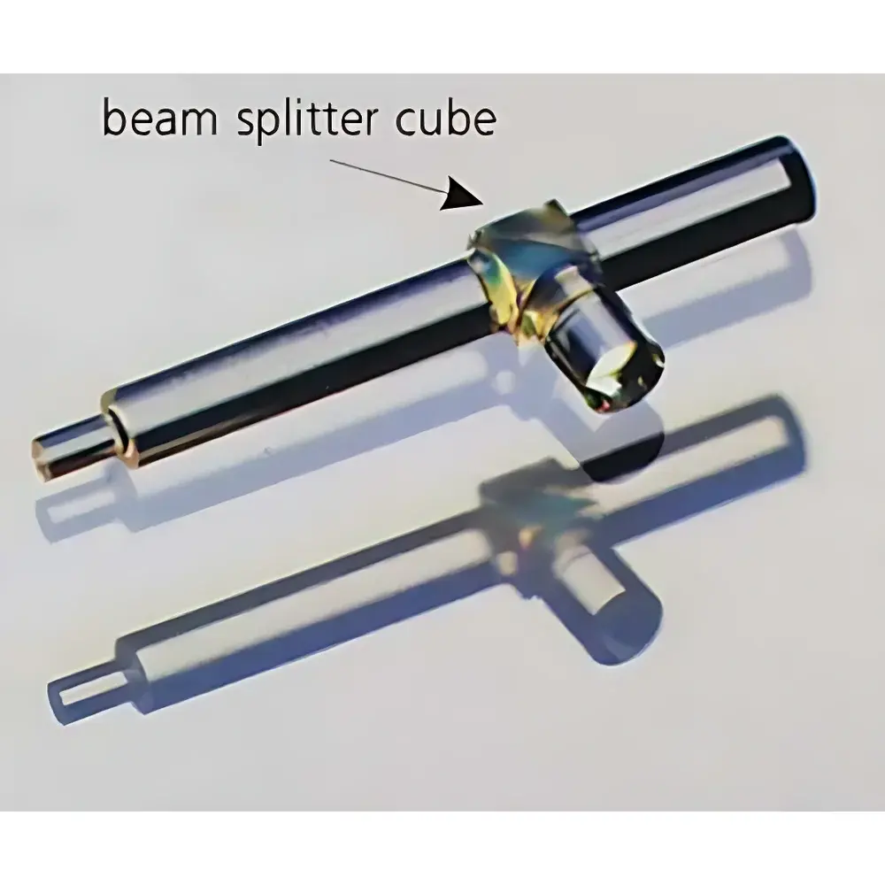
| Optical Customization | Prism-integrated beam steering, fiber/fiber bundle coupling, AR-coating (custom V-coat or broadband), dichroic/polarizing/non-polarizing beamsplitters, stainless-steel hypodermic tube housing |
| Compliance | Designed for ISO 13485-aligned biomedical research environments |
Overview
The GRINTECH GRIN Lens-Based In Vivo Confocal Microscopy and Optical Coherence Tomography (OCT) Probe System is a miniaturized, high-numerical-aperture optical imaging platform engineered for intravital, minimally invasive visualization of biological tissues at cellular and subcellular resolution. Leveraging gradient-index (GRIN) lens technology, this probe integrates dual-modal functionality—confocal laser scanning microscopy (CLSM) and spectral-domain OCT—within a single compact distal optics assembly. Unlike conventional objective-based systems, GRIN lenses eliminate the need for complex relay optics by exploiting radially varying refractive index profiles to focus and relay light through millimeter-scale diameters. This enables deep-tissue access in constrained anatomical spaces such as murine colonic lumen, pancreatic ducts, or cerebral ventricles, while maintaining diffraction-limited performance and minimal optical aberration. The system operates with solid-state laser excitation sources (typically 488 nm, 561 nm, or 640 nm for confocal fluorescence; 1310 nm or 1550 nm swept-source or broadband for OCT), and supports both point-scanning and line-field configurations depending on host instrument architecture.
Key Features
- Modular GRIN lens assemblies available in four standardized outer diameters: 0.5 mm, 1.0 mm, 1.8 mm, and 2.0 mm—enabling selection based on anatomical access requirements and mechanical stability constraints.
- Integrated prism-based beam redirection for 90° or adjustable-angle lateral illumination and detection, facilitating side-viewing endoscopic geometry without external mirror articulation.
- Fiber-optic or fiber-bundle coupling capability for seamless integration with commercial confocal microscopes (e.g., Zeiss LSM, Nikon A1R) and OCT engines (e.g., Thorlabs GAN, Michelson Diagnostics Callisto).
- Custom optical design services include ray-traced optimization of working distance, field-of-view, depth-of-field, and chromatic correction across dual-wavelength bands (visible + near-infrared).
- Optional anti-reflection coatings tailored to specific laser lines or broadband OCT spectra (e.g., R<0.25% @ 488/561/640 nm; R<0.3% @ 1310±50 nm); dichroic, polarizing, or non-polarizing beamsplitter integration within the probe housing.
- Stainless-steel hypodermic tubing encapsulation ensures biocompatibility, mechanical rigidity, and compatibility with standard stereotactic or endoscopic insertion protocols under GLP-compliant preclinical studies.
Sample Compatibility & Compliance
This probe system is validated for use in live animal models including murine colon, brain, kidney, and trachea—demonstrated in peer-reviewed studies using transgenic fluorescent reporters (e.g., Lgr5-EGFP, Rosa26-tdTomato). It meets dimensional and material specifications required for IACUC-approved intravital imaging protocols. While not a medical device per se, its construction adheres to ISO 10993-5 (cytotoxicity) and ISO 10993-10 (irritation/sensitization) testing guidelines when stainless-steel housings and USP Class VI-certified adhesives are specified. All optical components comply with RoHS Directive 2011/65/EU and REACH Regulation (EC) No. 1907/2006. Integration into regulated workflows requires validation of the full imaging chain—including laser safety classification (IEC 60825-1), data integrity controls, and audit trail generation per 21 CFR Part 11 when paired with compliant acquisition software.
Software & Data Management
The probe itself is hardware-agnostic and does not include embedded firmware or proprietary drivers. It interfaces transparently with industry-standard acquisition platforms including LabVIEW-based custom control systems, MATLAB Image Acquisition Toolbox, and commercial OEM SDKs (e.g., Thorlabs OCT SDK, Bruker Prairie View). Synchronized dual-modality acquisition requires time-stamped trigger alignment between confocal frame clocks and OCT A-scan triggers—a capability supported via TTL I/O ports on compatible controllers. Metadata embedding (e.g., lens NA, working distance, calibration coefficients) follows MIAME/MINSEQ-compliant schema when exported to OME-TIFF or HDF5 formats. For longitudinal studies, DICOM-SR extensions may be implemented for structured reporting of imaging parameters in preclinical PACS environments.
Applications
- Intravital monitoring of epithelial barrier integrity and immune cell trafficking in murine colitis models.
- Real-time assessment of tumor margin delineation during experimental endoscopic resection procedures.
- Dynamic imaging of neuronal calcium activity combined with structural OCT angiography in thinned-skull cortical preparations.
- Quantitative morphometric analysis of crypt architecture and goblet cell distribution in intestinal organoids cultured within microfluidic chips.
- Validation of nanoparticle biodistribution and clearance kinetics using co-registered fluorescence intensity and OCT backscatter contrast.
FAQ
Is this probe compatible with commercial confocal microscopes?
Yes—provided the host system supports external galvo or resonant scanner synchronization and offers fiber-coupled input/output ports. Integration typically requires collimation optics and achromatic focusing relays matched to the GRIN lens focal properties.
Can the probe be sterilized for repeated in vivo use?
Stainless-steel-housed variants support autoclaving at 121°C for 20 minutes (validated per ISO 17664); however, GRIN lens elements and fiber terminations require low-temperature alternatives such as ethylene oxide (EtO) or hydrogen peroxide plasma sterilization.
What is the typical working distance and field of view for the 1.0 mm diameter probe?
Standard configuration yields ~150 µm working distance and ~120 µm lateral field of view at 488 nm excitation; exact values depend on GRIN pitch length, numerical aperture, and relay optics design—available upon request with Zemax® simulation reports.
Do you provide optical calibration standards or reference phantoms?
GRINTECH offers NIST-traceable USAF 1951 resolution targets and scattering phantoms (e.g., Intralipid®-based tissue-simulating gels) for quantitative PSF characterization and OCT sensitivity verification.
Is technical support available for system integration and alignment?
Yes—application engineers provide remote assistance for optical alignment, signal-to-noise optimization, and modality synchronization; on-site support is available under service-level agreement (SLA) terms.


