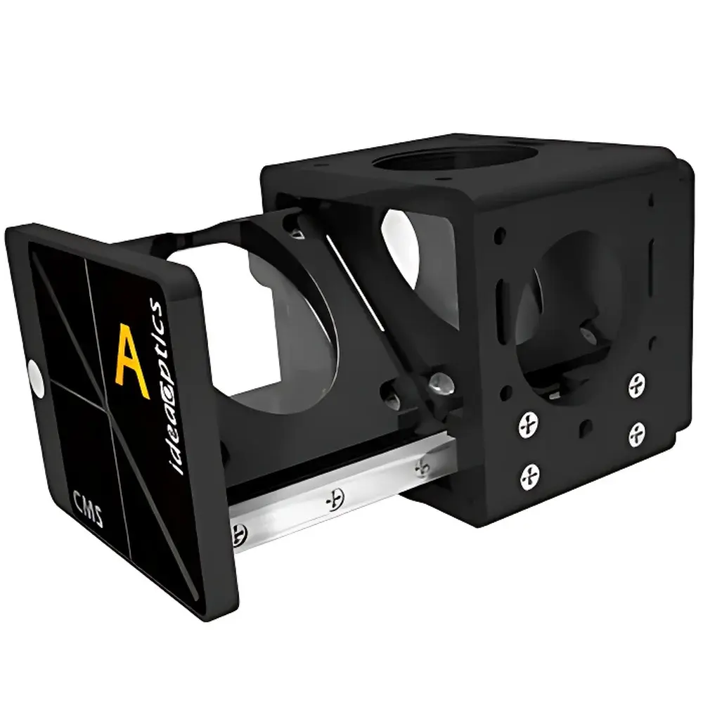
IdeaOptics CMS Microscope Spectral Expansion Port
| Brand | IdeaOptics |
|---|---|
| Origin | Shanghai, China |
| Manufacturer Type | OEM Manufacturer |
| Product Category | Optical Component |
| Model | CMS |
| Spectral Range | 200–2500 nm |
| Spatial Resolution (with FIB-M fiber) | ≤1 μm |
| Compatibility | Universal microscope mounting (custom interface provided) |
| Optical Functionality | Transmission/Reflection, Fluorescence, Raman spectroscopy |
| Beam Path Design | Dual-mode optical path (simultaneous imaging & spectral acquisition, micro-area light delivery) |
Overview
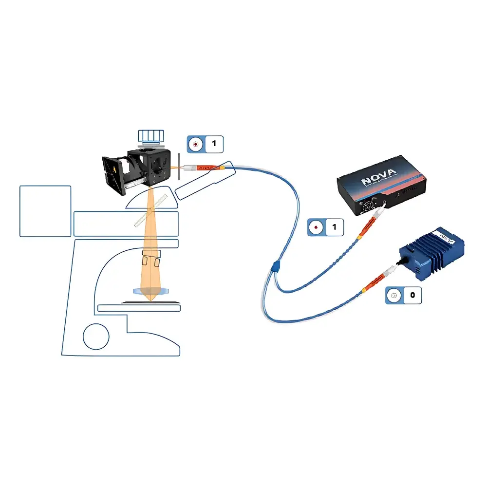
The IdeaOptics CMS Microscope Spectral Expansion Port is an engineered optical interface module designed to integrate broadband spectral analysis capabilities—spanning ultraviolet to short-wave infrared (200–2500 nm)—directly into standard upright and inverted optical microscopes. Unlike conventional spectrometer couplers that rely on external fiber coupling or bulk beam extraction, the CMS implements a dual-path internal optical architecture: one path preserves real-time visual observation through the eyepiece or camera port, while the second path directs wavelength-resolved light—selected via interchangeable dichroic mirrors and bandpass filters—to an external spectrometer. This co-registered “see-and-measure” functionality enables spatially resolved microspectroscopy without sacrificing morphological context. The CMS operates on principles of collimated beam routing and high-fidelity spectral throughput preservation, making it suitable for applications demanding strict photometric linearity, minimal polarization dependence, and low stray-light contribution—particularly in research-grade photonic material characterization.
Key Features
- Universal microscope integration: Compact form factor (≤85 mm × 65 mm × 42 mm) with mechanical adaptability to major commercial platforms (Olympus BX, Nikon Eclipse, Zeiss Axio series); custom mechanical and optical interface design provided at no additional cost.
- Sub-micron spatial resolution: When coupled with a 1-μm-core FIB-M (Fused Silica Imaging Bundle) optical fiber, the CMS supports spectral acquisition from defined regions as small as 1 μm in diameter—enabling diffraction-limited micro-area spectroscopy under 100× objective magnification.
- Full UV–SWIR spectral coverage: Internal modular dichroic mount accommodates up to three user-replaceable beamsplitters and filter sets, supporting seamless switching between transmission/reflection, fluorescence, and Raman configurations across 200–2500 nm.
- Dual-path optical design: Simultaneous optical imaging and spectral signal extraction ensure precise correlation between morphological features and spectral response—critical for heterogeneous sample analysis and process validation.
- Low-loss optical train: All internal optics utilize λ/10 surface flatness fused silica substrates with MgF₂ anti-reflection coatings (R<0.5% per surface, 200–2500 nm), minimizing insertion loss and spectral distortion.
Sample Compatibility & Compliance
The CMS is routinely deployed in GLP-compliant laboratories for characterization of photonic and microelectromechanical systems where traceable, repeatable microspectral data are required. It supports non-destructive, ambient-condition measurement of solid-state samples including self-assembled photonic crystals, MEMS cantilevers, semiconductor nanowires, plasmonic metasurfaces, and thin-film optoelectronic devices. While the CMS itself is not a standalone measuring instrument, its optical interface design conforms to ISO 10110-7 (optical component surface quality) and complies with mechanical interface standards referenced in ASTM E2821–22 (“Standard Guide for Microspectrophotometry”). When integrated into a full system with NIST-traceable spectrometers and calibrated light sources, measurement uncertainty budgets align with ISO/IEC 17025 requirements for spectral radiance and reflectance.
Software & Data Management
The CMS requires no proprietary control software; it functions transparently within existing microscope control environments (e.g., Olympus cellSens, Nikon NIS-Elements, Zeiss ZEN). Spectral data acquisition is managed via third-party spectrometer SDKs (e.g., OceanInsight OceanView, Hamamatsu HCImage, Andor Solis). For traceability and audit readiness, users may configure metadata tagging—including microscope objective ID, magnification, excitation wavelength, filter set ID, and CMS optical path mode—directly in the spectrometer acquisition software. The dual-path architecture inherently supports time-synchronized image–spectrum capture, enabling pixel-to-spectrum mapping workflows compatible with HDF5-based data storage frameworks used in FAIR-compliant materials informatics pipelines.
Applications
- Photonic crystal domain mapping: Resolving local stopband shifts across individual micro-domains (<5 μm) in self-assembled opal structures using reflectance microspectroscopy at 1–2 nm spectral resolution.
- MEMS optical response validation: Measuring position-dependent transmittance/reflectance spectra from sub-10 μm actuator elements under electrostatic bias, supporting design verification against FDTD simulation outputs.
- Nanowire fluorescence fingerprinting: Acquiring excitation-emission matrices (EEMs) from single CdSe/ZnS core-shell nanowires with <1.5 nm spectral bandwidth and <0.3% peak intensity repeatability over 100-cycle thermal cycling.
- Surface-enhanced Raman substrate screening: Rapid assessment of SERS enhancement uniformity across lithographically patterned Au nanoantenna arrays via 785 nm excitation and 5 cm⁻¹ spectral resolution.
FAQ
Is the CMS compatible with confocal laser scanning microscopes (CLSM)?
Yes—the CMS can be integrated into the side-port or rear-port output of most CLSM platforms, provided the optical path allows insertion of a 1-inch diameter collimated beam. Custom kinematic mounts are available upon request.
Can the CMS be used for time-resolved photoluminescence (TRPL)?
It supports TRPL when paired with a streak camera or time-correlated single-photon counting (TCSPC) spectrometer; however, temporal resolution is governed by the external detection system—not the CMS optical path.
Does the CMS introduce chromatic aberration or polarization artifacts?
No—its all-fused-silica optical train and achromatic beam-splitting design maintain wavefront fidelity across the full 200–2500 nm range; polarization extinction ratio exceeds 1000:1 in reflection mode.
What maintenance is required?
None beyond routine dust prevention; all optical surfaces are sealed and coated for environmental stability. No alignment screws or user-serviceable components are present.
Is calibration documentation provided?
A factory spectral throughput certificate (measured against NIST SRM 2036) and mechanical interface dimensional report are included with each unit; full system calibration must be performed by the end-user in situ.


