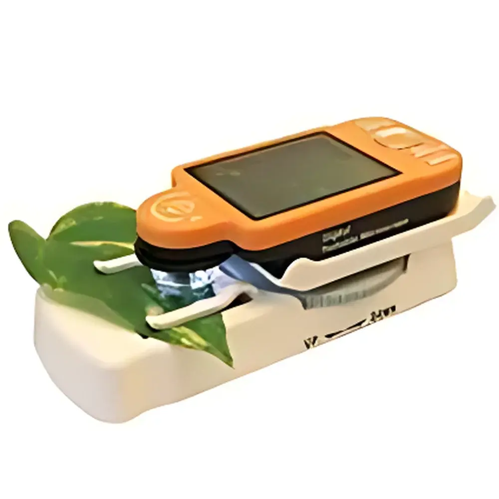
IPM CAM2 Portable Plant Leaf Imaging System with Integrated Digital Microscope
| Origin | USA |
|---|---|
| Manufacturer Type | Authorized Distributor |
| Import Status | Imported |
| Model | IPM CAM2 |
| Pricing | Available Upon Request |
| Power Supply | USB-powered or Rechargeable Li-ion Battery |
| Light Source | 4 Adjustable-Brightness High-Intensity LED Lamps |
| Sensor Resolution | 2 MP (1600 × 1200 interpolated), Native 640 × 480 |
| Optical Zoom | 10× or 40× |
| Digital Zoom | Up to 200× |
| Field of View (FOV) | 40× = 8 mm × 10 mm |
| Working Distance | Fixed focus, optimized for leaf surface imaging (0.5–2 cm) |
| Output Interface | USB 2.0 |
| Display | Built-in 3.5″ TFT LCD |
| Image/Video Format | JPEG, BMP, AVI |
| Data Annotation | On-screen measurement tools and timestamped labeling |
Overview
The IPM CAM2 Portable Plant Leaf Imaging System is a field-deployable, integrated digital microscopy platform engineered for non-destructive, high-fidelity documentation of leaf surface morphology, stomatal architecture, epidermal cell patterning, and early-stage biotic/abiotic stress indicators. Unlike conventional benchtop fluorescence or confocal systems, the IPM CAM2 operates on a simplified coaxial illumination and fixed-focus optical design grounded in brightfield microscopy principles—eliminating complex alignment procedures while preserving spatial fidelity at biologically relevant magnifications (40×–140×). Its compact, ruggedized housing (IP54-rated against dust and light splashing) enables rapid deployment across diverse ecological settings—from greenhouse phenotyping trials to remote forest understory surveys—without reliance on external power grids or vibration-isolated tables. The system captures real-time video and stills directly to host computers or onboard storage, supporting time-series monitoring of dynamic physiological responses such as stomatal aperture changes, trichome density shifts, or lesion development under controlled or ambient conditions.
Key Features
- Field-optimized optical architecture: Fixed-focus lens assembly calibrated for leaf surface imaging at working distances of 0.5–2 cm, minimizing parallax error during handheld or tripod-assisted operation.
- Dual-mode illumination: Four independently controllable high-CRI LED sources provide uniform, shadow-free illumination; brightness adjustment enables contrast optimization for waxy cuticles, anthocyanin-rich tissues, or chlorotic zones.
- Real-time visualization and annotation: Integrated 3.5″ LCD screen supports immediate image review, manual ROI selection, distance calibration, and timestamped markup—critical for GLP-compliant field logbooks.
- USB 2.0 plug-and-play interface: Compatible with Windows, macOS, and Linux without proprietary drivers; exports standardized JPEG/BMP stills and AVI video streams for downstream analysis in ImageJ, FIJI, or custom Python-based segmentation pipelines.
- Battery-resilient operation: Rechargeable Li-ion battery sustains >4 hours of continuous imaging at 40×; USB bus power provides uninterrupted operation when tethered to laptops or portable power banks.
Sample Compatibility & Compliance
The IPM CAM2 is validated for intact, unsectioned leaf samples from angiosperms, gymnosperms, and ferns—including species with high surface reflectance (e.g., Eucalyptus, Populus) or delicate epidermal layers (e.g., Arabidopsis thaliana, rice seedlings). No mounting media, vacuum desiccation, or conductive coating is required. The system complies with ISO 17025 general requirements for competence of testing and calibration laboratories when used within documented SOPs. Image metadata (exposure time, magnification, illumination intensity, timestamp) are embedded in EXIF headers, satisfying traceability requirements under FDA 21 CFR Part 11 for regulated agricultural research and seed certification workflows.
Software & Data Management
The included IPM Capture Suite (v3.2+) provides lossless image acquisition, pixel-to-micron calibration via stage micrometer reference, and batch export with user-defined naming conventions (e.g., “PlotID_Date_Strain_Treatment”). All annotations—including scale bars, region-of-interest polygons, and measurement overlays—are stored as XML sidecar files, ensuring audit trail integrity. Raw data exports conform to FAIR principles (Findable, Accessible, Interoperable, Reusable), facilitating integration with LIMS platforms or cloud-based phenotyping repositories such as BreedBase or CyVerse. Optional API access enables automated triggering via GPIO or serial command for synchronized multi-sensor deployments (e.g., concurrent chlorophyll fluorescence and structural imaging).
Applications
- Stomatal conductance proxy quantification via aperture width/density mapping under drought or CO2 enrichment treatments.
- Rapid screening of foliar disease symptoms (e.g., powdery mildew hyphae, rust pustules) in breeding nurseries.
- Time-lapse documentation of herbicide phytotoxicity progression or nutrient deficiency morphotypes.
- Validation of hyperspectral or thermal imaging outputs through ground-truth microstructural correlation.
- Education and outreach: Real-time classroom demonstration of leaf anatomy without slide preparation or staining.
FAQ
Is the IPM CAM2 suitable for fluorescence imaging?
No—the system utilizes brightfield illumination only and lacks excitation/emission filter sets or UV-LED sources required for chlorophyll a or GFP autofluorescence detection. For fluorescence applications, consider the companion IPM FLUO series.
Can it be mounted on a stereo microscope stand?
Yes—standard 1/4″-20 threaded base allows secure attachment to modular lab stands or custom field tripods with adjustable height and tilt.
What is the minimum resolvable feature size at 140×?
Based on Nyquist sampling theory and native sensor resolution, the practical limit is ~3.5 µm under optimal lighting and focus—sufficient to resolve individual stomatal guard cells in most crop species.
Does it support live measurement units (e.g., µm, mm) during acquisition?
Yes—calibration using a certified stage micrometer enables real-time on-screen dimensional readouts with ±2% uncertainty across the FOV.
Is firmware upgradable in the field?
Yes—USB-based firmware updates preserve backward compatibility with existing capture software and add support for new file formats or annotation tools per annual release cycle.


