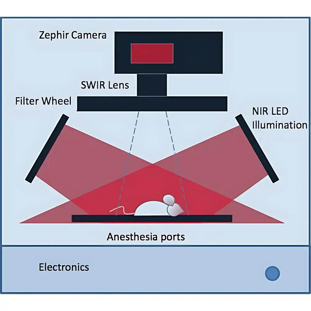
IR VIVO NIR-II Multispectral In Vivo Imaging System
| Origin | Czech Republic |
|---|---|
| Manufacturer Type | Authorized Distributor |
| Origin Category | Imported Instrument |
| Model | IR VIVO NIR-II Multispectral In Vivo Imaging System |
| Price Range | USD 13,500 – 27,000 |
Overview
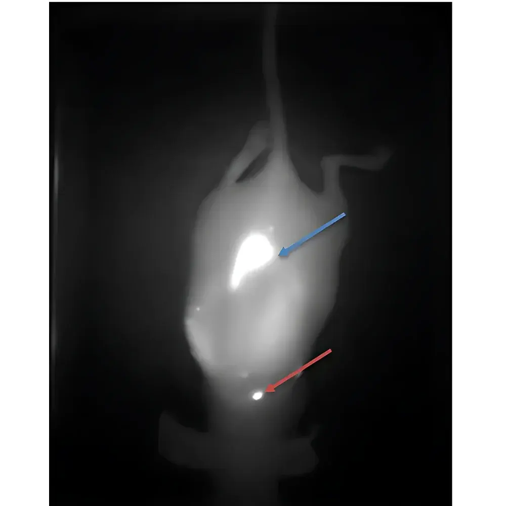
The IR VIVO NIR-II Multispectral In Vivo Imaging System is a high-performance preclinical optical imaging platform engineered for non-invasive, real-time visualization of physiological and pathological processes in live small animals. Operating within the second near-infrared window (NIR-II, 850–1600 nm), it leverages reduced photon scattering and minimal tissue autofluorescence to achieve superior penetration depth (>3 cm in soft tissue), sub-millimeter spatial resolution, and high temporal fidelity—enabling functional, dynamic, and quantitative imaging without ionizing radiation or exogenous contrast toxicity. Unlike conventional visible-light or first-window NIR-I systems, the IR VIVO system exploits the intrinsic optical advantages of the NIR-II band to resolve deep-tissue vascular architecture, tumor metabolism, inflammatory cell trafficking, and pharmacokinetic distribution with high anatomical-functional correlation. Its design complies with international standards for preclinical instrumentation used in GLP-compliant studies, including ISO 13485-aligned manufacturing traceability and compatibility with FDA 21 CFR Part 11–ready data acquisition workflows.
Key Features
- True NIR-II spectral operation (850–1600 nm) with tunable LED excitation sources (standard 780 nm and 810 nm; optional extended wavelengths)
- High-sensitivity InGaAs focal plane array detector (640 × 512 pixels, 15 µm pixel pitch) optimized for low-noise, high-dynamic-range signal capture
- Adjustable field-of-view (3.1 × 2.5 cm to 15.5 × 12.5 cm) with uniform illumination over 15.5 × 12.5 cm area
- Multi-spectral acquisition capability enabling spectral unmixing of endogenous chromophores (e.g., oxy-/deoxy-hemoglobin, water, lipids) and exogenous probes (e.g., IR-26, CH1055, LS-501)
- Sub-second temporal resolution supporting dynamic perfusion mapping, cardiac gating, and real-time drug biodistribution tracking
- Robust mechanical architecture with motorized zoom lens, precision stage control, and integrated thermal stabilization for long-duration acquisitions
Sample Compatibility & Compliance
The IR VIVO system supports longitudinal imaging of murine models (C57BL/6, BALB/c, nude, NSG), rat strains (Sprague-Dawley, Wistar), and ex vivo tissue specimens—including brain slices, tumor explants, and vascular grafts. It accommodates standard anesthesia interfaces (isoflurane-compatible nose cone and gas scavenging ports) and temperature-regulated animal beds (36.5 ± 0.3 °C). All hardware and firmware are designed to meet IEC 61000-6-3 (EMC emissions) and IEC 61000-6-2 (immunity) requirements. Data integrity protocols align with ALCOA+ principles (Attributable, Legible, Contemporaneous, Original, Accurate, Complete, Consistent, Enduring, Available), supporting audit readiness for regulatory submissions under USP , ISO/IEC 17025, and OECD GLP guidelines.
Software & Data Management
The proprietary IR VIVO Acquisition Suite provides intuitive, scriptable control of exposure time, gain, spectral binning, and stage positioning via a Windows-based GUI. Image processing modules include spectral deconvolution, ratiometric hemodynamic mapping (HbO₂/Hb), time-intensity curve fitting, and 3D surface reconstruction from multi-angle acquisitions. Raw data are stored in vendor-neutral HDF5 format with embedded metadata (acquisition timestamp, instrument calibration ID, animal ID, protocol version). The software supports DICOM export for cross-platform integration with PACS or MATLAB/Python-based analysis pipelines. Optional validation packages include IQ/OQ documentation templates and electronic audit trail logging compliant with 21 CFR Part 11 Subpart B.
Applications
- Preclinical oncology: Tumor angiogenesis quantification, metastatic nodules detection, and targeted probe validation
- Neurovascular research: Cerebral blood flow dynamics, blood-brain barrier permeability assessment, and stroke model progression monitoring
- Cardiovascular pharmacology: Myocardial perfusion imaging, atherosclerotic plaque characterization, and stent endothelialization tracking
- Immunology & inflammation: Lymph node drainage kinetics, macrophage polarization mapping, and cytokine reporter expression
- Toxicology & ADME: Real-time hepatic/renal clearance profiling and nanoparticle organ accumulation kinetics
- Metabolic phenotyping: Brown adipose tissue activation, glucose uptake dynamics, and mitochondrial redox state imaging
FAQ
What is the maximum achievable imaging depth in biological tissue using the IR VIVO system?
Typical penetration depth exceeds 3 cm in murine abdominal tissue and ~1.5 cm in intact rat brain under optimal spectral settings—approximately 10× greater than conventional visible-light imaging.
Does the system support quantitative spectral unmixing of multiple fluorophores?
Yes; the multispectral acquisition engine enables linear unmixing of up to six spectrally distinct probes simultaneously when calibrated against reference spectra.
Is the IR VIVO system compatible with existing laboratory infrastructure such as isoflurane anesthesia units?
Yes; it includes standardized gas inlet/outlet ports and a modular animal holder with integrated gas delivery channels and thermal feedback control.
Can raw image data be exported for third-party analysis (e.g., MATLAB, Python, ImageJ)?
All acquired datasets are saved in HDF5 format with full metadata, and open-source Python libraries (e.g., h5py, scikit-image) are fully supported for downstream processing.
What regulatory documentation is provided for GLP-compliant study use?
Standard delivery includes Factory Acceptance Test (FAT) report, Installation Qualification (IQ) checklist, and Operational Qualification (OQ) test protocols aligned with ISO/IEC 17025 Annex A. Custom validation support is available upon request.


