LaVision StrainMaster DVC Digital Volume Correlation System
| Brand | LaVision GmbH |
|---|---|
| Origin | Germany |
| Model | StrainMaster DVC |
| Application Domain | 3D Full-Field Internal Strain & Displacement Mapping |
| Imaging Modality Compatibility | X-ray CT, MRI, OCT, Optical Tomography |
| Technique Principle | Digital Volume Correlation (DVC) |
| Compliance Framework | Supports GLP/GMP-aligned data traceability and ASTM E2774–19 (Standard Guide for Digital Image Correlation) |
Overview
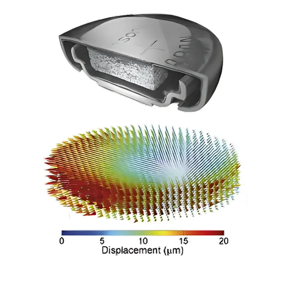
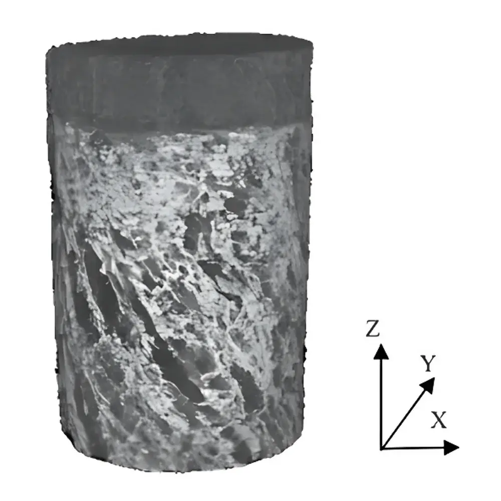
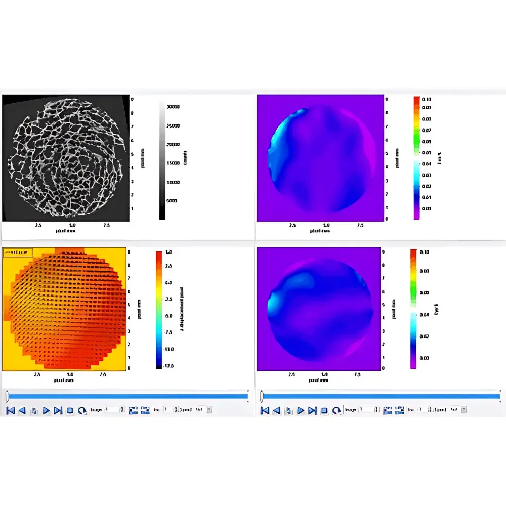
The LaVision StrainMaster DVC Digital Volume Correlation System is an advanced, non-invasive metrology platform engineered for quantitative, volumetric strain and displacement analysis in solid materials under mechanical, thermal, or electrochemical loading. Unlike surface-based techniques such as digital image correlation (DIC), StrainMaster DVC operates on registered 3D volumetric image stacks—typically acquired via laboratory or synchrotron-based X-ray computed tomography (X-ray CT), magnetic resonance imaging (MRI), optical coherence tomography (OCT), or laser-scanning confocal microscopy—to compute full-field 3D displacement vectors and derived strain tensors (εxx, εyy, εzz, γxy, γyz, γxz) at sub-voxel resolution. The system implements a robust, iterative cross-correlation algorithm applied across user-defined interrogation sub-volumes (IVs), leveraging inherent material heterogeneity—e.g., microstructural porosity, phase contrast, or intentionally introduced tracer particles—as the internal speckle pattern required for correlation stability. This enables direct quantification of internal deformation mechanisms including localized plasticity, crack tip fields, particle-matrix decohesion, and time-dependent viscoelastic relaxation—without requiring physical sensor integration or specimen sectioning.
Key Features
- Native support for multi-modal 3D image data: imports reconstructed volume stacks from X-ray CT (lab/synchrotron), MRI, OCT, and optical tomography systems in standard formats (e.g., TIFF stack, NIfTI, DICOM, MHD)
- Adaptive sub-volume sizing and overlap strategy to balance spatial resolution, noise sensitivity, and computational efficiency
- Robust motion compensation and rigid-body subtraction prior to deformation analysis, minimizing artifacts from stage drift or sample repositioning
- Comprehensive strain tensor derivation: includes Green-Lagrange, Euler-Almansi, and infinitesimal formulations with optional smoothing and outlier rejection filters
- GPU-accelerated correlation engine enabling scalable processing of volumes up to 2048³ voxels on workstation-class hardware
- Integrated uncertainty estimation module based on correlation peak sharpness, subset deformation gradient conditioning, and synthetic displacement field validation
Sample Compatibility & Compliance
StrainMaster DVC accommodates specimens ranging from millimeter-scale biomedical scaffolds to centimeter-scale composite coupons and battery electrodes. It requires no surface preparation or coating—making it ideal for in situ/operando experiments inside environmental chambers, mechanical testers, or electrochemical cells. The system supports compliance-critical workflows: raw image metadata (including acquisition parameters, timestamps, and scanner calibration coefficients) is preserved throughout processing; all intermediate and final results are stored with embedded audit trails; and software versioning, user authentication, and parameter logging align with principles outlined in ASTM E2774–19 and ISO/IEC 17025:2017 for measurement validity. While not FDA-certified as standalone medical device software, its architecture meets foundational requirements for Part 11–compliant environments when deployed with validated IT infrastructure and procedural controls.
Software & Data Management
The StrainMaster DVC software suite provides a modular, scriptable environment built on Qt and Python (via PySide and NumPy/SciPy). Users define workflows through a visual pipeline editor or programmatically using the StrainMaster Python API. Processed results—including displacement fields, strain maps, and principal strain orientations—are exportable in HDF5, VTK, or CSV formats for downstream finite element model validation (e.g., ABAQUS, ANSYS) or statistical microstructure–property analysis. Built-in visualization tools support interactive 3D slicing, vector glyphs, iso-surface rendering, and temporal sequence playback. All processing steps are reproducible via JSON-based protocol files, enabling full method transfer between laboratories and long-term archival integrity.
Applications
- Mechanical characterization of cellular solids (e.g., auxetic foams, trabecular bone analogs) under uniaxial/biaxial compression
- In situ tracking of lithiation-induced dilation and fracture nucleation in Li-ion battery electrodes during cycling (as demonstrated in Advanced Energy Materials, Eastwood et al.)
- Quantitative assessment of scaffold-host tissue integration mechanics in cartilage repair implants (micro-CT–based DVC, University of Portsmouth)
- Damage initiation mapping in fiber-reinforced polymer composites subjected to fatigue or impact loading
- Thermo-mechanical strain evolution in thermal barrier coatings and additive-manufactured metallic lattices
- Microscale deformation partitioning in multiphase alloys and metal matrix composites
FAQ
What image resolution is required for reliable DVC measurements?
Spatial resolution must resolve at least 3–5 pixels per characteristic feature size (e.g., pore diameter or particle spacing) to ensure sufficient contrast gradient for correlation. Typical lab CT systems operating at ≤5 µm voxel size yield robust results for most engineering materials.
Can StrainMaster DVC process time-lapse or 4D datasets?
Yes—the software natively handles sequential volume stacks acquired over time or load steps, enabling transient strain evolution analysis with automated registration and differential correlation.
Is a physical speckle pattern required inside the sample?
No. StrainMaster DVC relies on intrinsic structural heterogeneity (e.g., pores, second-phase particles, density gradients) as the natural volume speckle. Artificial tracers (e.g., tungsten nanoparticles in polymers) may be introduced only when native contrast is insufficient.
Does the system support batch processing of multiple samples?
Yes—via command-line interface (CLI) mode and Python scripting, enabling unattended execution of standardized protocols across large experimental series.
How is measurement uncertainty quantified?
Uncertainty is estimated per voxel using normalized cross-correlation confidence metrics, deformation gradient condition number analysis, and optional synthetic ground-truth validation with known displacement fields.

")

