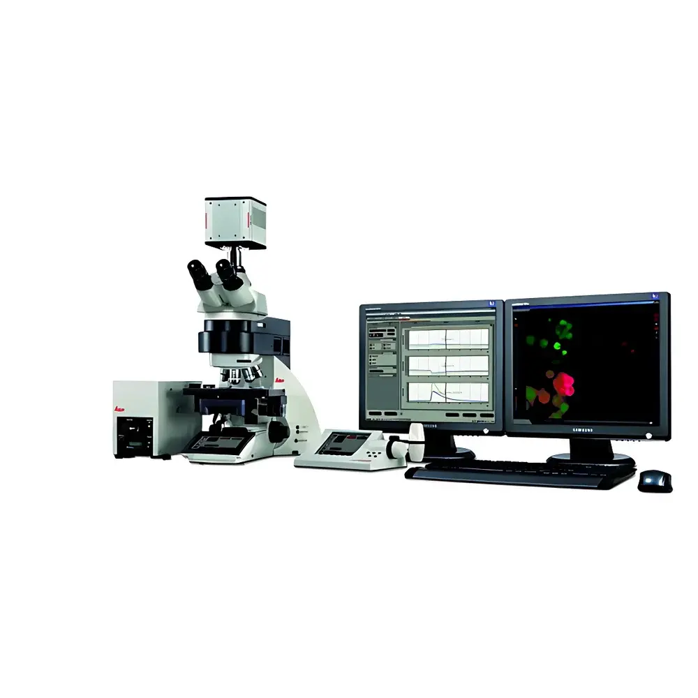
Leica AF6000 Upright Fluorescence Microscopy System
| Brand | Leica |
|---|---|
| Origin | Germany |
| Model | AF6000 |
| Instrument Type | Upright Fluorescence Microscope |
| Excitation Source | Laser-based Illumination System |
| Application Platform | Integrated Widefield & Fluorescence Imaging for Live-Cell and Fixed-Sample Analysis |
| Software Suite | Leica Application Suite X (LAS X) with Advanced Quantitative Modules |
Overview
The Leica AF6000 is an upright fluorescence microscopy system engineered for high-precision, multi-modal imaging in life science research laboratories. Unlike inverted configurations optimized for cell culture dishes, the AF6000’s upright architecture supports a broad range of specimen formats—including glass-mounted tissue sections, whole-mount embryos, thick histological slices, and live specimens in specialized chambers—while maintaining optical stability and ergonomic accessibility. Its core design integrates widefield epifluorescence illumination with laser-driven excitation pathways, enabling simultaneous or sequential acquisition across multiple fluorophores with minimal crosstalk. The system operates on a rigid, vibration-damped optical bench and employs infinity-corrected optics compatible with Leica’s full line of HCX and PL FLUOTAR objectives (10× to 100×, NA up to 1.4). Designed explicitly for quantitative fluorescence applications, the AF6000 delivers consistent photon collection efficiency, low autofluorescence background, and thermal stabilization of critical optical components—key requirements for time-lapse, Z-stack, and deconvolution workflows compliant with GLP and ISO/IEC 17025 laboratory practices.
Key Features
- Modular upright platform supporting interchangeable fluorescence filter cubes, motorized filter turrets, and automated shutter control for precise exposure timing.
- Laser-based excitation system with selectable wavelengths (e.g., 405 nm, 488 nm, 561 nm, 640 nm), integrated acousto-optic tunable filters (AOTFs) or dichroic mirror sets for spectral flexibility and rapid channel switching.
- Motorized XYZ stage with sub-micron repeatability and programmable coordinate memory for multi-position time-lapse experiments across standard microscope slides or custom sample holders.
- High-sensitivity sCMOS or EMCCD camera integration with hardware-triggered synchronization for low-noise, high-dynamic-range image capture at frame rates up to 30 fps (full resolution).
- Automated Z-focus maintenance via hardware-based focus drift compensation (e.g., Leica Adaptive Focus Control), essential for extended live-cell imaging sessions exceeding several hours.
- Standard compliance with DIC, phase contrast, and brightfield imaging modes—enabling correlative structural and functional analysis without repositioning specimens.
Sample Compatibility & Compliance
The AF6000 accommodates diverse biological specimens: paraffin-embedded and frozen tissue sections (1–100 µm thickness), cleared whole organs (e.g., CLARITY, iDISCO), live zebrafish or C. elegans embryos in agarose mounts, and acute brain slices in perfusion chambers. Its open-stage design permits integration of environmental control units (temperature, CO₂, humidity) for long-term viability during in vivo imaging. All optical paths meet ISO 10934-1 (microscope terminology) and ISO 9335 (fluorescence microscope performance testing) standards. Data acquisition workflows support audit trails, electronic signatures, and metadata embedding per FDA 21 CFR Part 11 requirements when operated with LAS X software in validated configurations.
Software & Data Management
Leica Application Suite X (LAS X) serves as the unified control and analysis environment. It provides real-time image preview, multi-dimensional acquisition scripting (time, position, Z, channel), and embedded GPU-accelerated deconvolution using constrained iterative algorithms (e.g., Classic or Wiener methods). Quantitative modules include FRET ratio imaging with acceptor photobleaching and sensitized emission correction, co-localization analysis (Pearson’s coefficient, Mander’s overlap), object-based segmentation with machine learning-assisted training (LAS X Navigator), and multi-well plate mapping for high-content screening. Raw data is stored in TIFF or Leica’s proprietary LIF format, with optional DICOM export for cross-platform archival. All processing steps are logged with timestamps, user IDs, and parameter sets—ensuring traceability for regulatory submissions.
Applications
- Long-term time-lapse imaging of neuronal development, mitotic progression, or organelle dynamics in primary neurons or stem-cell-derived organoids.
- Quantitative spatial analysis of protein co-localization in fixed tumor biopsies or subcellular trafficking assays.
- FRET-based biosensor readouts for intracellular Ca²⁺, cAMP, or kinase activity in transfected mammalian cells.
- 3D reconstruction of embryonic patterning via automated Z-stack acquisition followed by volume rendering and surface modeling.
- Multi-site screening of compound effects across 96- or 384-well plates using standardized exposure and focus protocols.
- Correlative light microscopy (CLM) workflows aligned with subsequent electron microscopy (EM) grid mapping.
FAQ
Is the AF6000 compatible with third-party cameras or detectors?
Yes—the system supports industry-standard Camera Link and USB3 Vision interfaces, enabling integration with select sCMOS, EMCCD, and scientific CMOS cameras from vendors such as Hamamatsu, Andor, and Photometrics, provided firmware and driver compatibility is verified through Leica’s certified hardware list.
Does the AF6000 support super-resolution techniques like STED or SIM?
No—the AF6000 is a widefield fluorescence platform; it does not incorporate stimulated emission depletion lasers or structured illumination hardware. For super-resolution, Leica recommends the TCS SP8 STED or SR GSD 3D systems.
Can LAS X software be deployed in networked, multi-user environments?
Yes—LAS X supports concurrent licensing models and centralized configuration management via Leica’s LAS X Server, enabling role-based access control and shared instrument scheduling across departmental networks.
What environmental controls can be added for live-cell imaging?
Leica-certified incubation chambers (e.g., Life Science Stage Top Incubator) provide precise regulation of temperature (20–42 °C ±0.1 °C), CO₂ (0–20%), and humidity (up to 95% RH), all controllable via LAS X GUI with real-time feedback logging.
Is deconvolution processing performed in real time or post-acquisition?
Both options are available: preview deconvolution runs on GPU during acquisition for immediate focus assessment, while full-resolution batch deconvolution is executed offline with configurable iteration limits and regularization parameters.
Related Products


