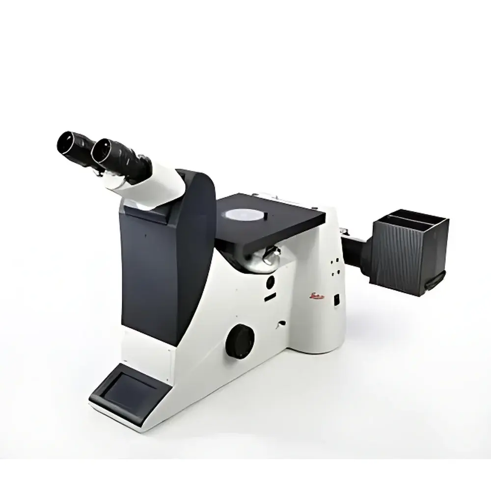
Leica DMI3000 M Inverted Manual Research-Grade Metallurgical Microscope
| Brand | Leica |
|---|---|
| Origin | Germany |
| Model | DMI3000 M |
| Type | Inverted Metallurgical Microscope |
| Optical Design | Achromatic-Apochromatic Corrected |
| Illumination | Reflected Light (Epi-Illumination) |
| Standard Contrast Modes | Brightfield, Darkfield, Oblique Illumination |
| Optional Contrast | Differential Interference Contrast (DIC) |
| Objective Mount | Quintuple Nosepiece |
| Stage | Mechanical XY Translation Stage |
| Low-Magnification Objective | 1.25× (for Wide-Field Overview) |
| Compliance | Designed to Support ISO 9001-Qualified Inspection Processes |
| Software Interface | Compatible with Leica Application Suite (LAS) X for Image Capture and Documentation |
Overview
The Leica DMI3000 M is a manually operated, inverted research-grade metallurgical microscope engineered for rigorous industrial and materials science applications. Unlike upright configurations, its inverted optical architecture positions the objective lenses beneath the specimen stage—enabling direct observation of large, heavy, or container-bound samples such as metallographic mounts, semiconductor wafers, castings, welds, and coated substrates without reorientation or disassembly. The system employs high-precision epi-illumination optics with coaxial reflected-light pathways, delivering exceptional depth of field, edge-to-edge resolution, and chromatic fidelity across magnifications. Its optical train integrates apochromatic correction for visible wavelengths and is optimized for high numerical aperture (NA) objectives, ensuring diffraction-limited performance in both brightfield and contrast-enhanced modalities. Designed for reproducible metrology-grade documentation, the DMI3000 M serves as a foundational platform for failure analysis, microstructure characterization, coating thickness evaluation, and process validation in R&D laboratories and QC environments compliant with ISO/IEC 17025 and ASTM E3–22 standards.
Key Features
- Inverted configuration with rigid, vibration-damped base and precision-machined optical column—optimized for stability during extended manual operation and high-magnification imaging.
- Quintuple nosepiece accommodating up to five high-NA metallurgical objectives (e.g., 5×, 10×, 20×, 50×, 100× oil), enabling rapid, repeatable magnification switching without refocusing.
- Dedicated oblique illumination module providing adjustable off-axis lighting—generating pseudo-DIC-like surface relief perception for topographic assessment of polished, etched, or uncoated metallic specimens where conventional contrast fails.
- Integrated brightfield/darkfield selector lever and modular filter turret—supporting seamless transition between standard metallurgical contrast modes without realignment.
- Mechanical XY stage with vernier-scaled translation (76 × 52 mm travel range) and specimen holder compatibility for standard 30 mm and 50 mm metallographic mounts.
- 1.25× wide-field objective included as standard—delivering panoramic overview imaging at low magnification for rapid defect localization and contextual mapping prior to high-resolution analysis.
Sample Compatibility & Compliance
The DMI3000 M accommodates specimens up to 120 mm in height and 20 kg in mass, including bulk castings, heat-treated components, PCB assemblies, and encapsulated electronic packages. Its open-stage design permits integration of thermal stages, hardness testers, or micro-manipulators for correlative analysis. All optical components meet Leica’s DIN-compliant manufacturing tolerances and are certified for long-term stability under continuous industrial use. The system supports documentation workflows aligned with GLP and GMP requirements—including audit-trail-capable image metadata capture when paired with LAS X software. While the base instrument operates without regulatory certification, its optical performance and mechanical repeatability conform to measurement uncertainty guidelines referenced in ISO 10772:2020 (Metallographic examination) and ASTM E1245–21 (Quantitative metallography).
Software & Data Management
The microscope is fully compatible with Leica Application Suite (LAS) X—a modular, FDA 21 CFR Part 11–ready imaging platform supporting calibrated measurement, multi-channel fluorescence overlay (when equipped with optional filters), time-lapse acquisition, and structured reporting. LAS X enables DIC vector alignment calibration, automatic focus mapping, and export of TIFF/PDF reports with embedded scale bars, operator ID, timestamp, and objective metadata. Raw image data preserves 16-bit dynamic range and EXIF-compliant acquisition parameters—ensuring traceability for internal audits or third-party accreditation assessments. No proprietary cloud services or mandatory subscriptions are required; local deployment on Windows-based workstations ensures full data sovereignty.
Applications
- Metallurgical quality assurance: grain size analysis per ASTM E112, inclusion rating per ASTM E45, porosity quantification in aluminum die-castings.
- Failure analysis: crack propagation path identification, intergranular corrosion assessment, solder joint integrity inspection in automotive electronics.
- R&D process development: heat treatment optimization, diffusion layer thickness measurement, thin-film adhesion evaluation via scratch testing correlation.
- Coating and plating verification: thickness uniformity mapping, interface delamination detection, anodized layer pore structure analysis.
- Geological and ceramic material characterization: phase distribution in sintered composites, refractory grain boundary analysis, slag inclusion morphology.
FAQ
Is motorized focusing or automated stage control available on the DMI3000 M?
No—the DMI3000 M is strictly manual in operation. Motorized Z-drive, encoded stage, or autofocus modules are exclusive to the DMI6000/DMI8 series. This design prioritizes mechanical robustness, cost efficiency, and service longevity for high-volume routine inspection.
Can DIC be added post-purchase?
Yes—DIC functionality requires installation of a Nomarski prism set in the condenser and matching prism-equipped objectives. Leica offers factory-certified retrofit kits with optical alignment verification and recalibration documentation.
What is the maximum usable magnification with the 1.25× objective?
When combined with a 10× eyepiece, the 1.25× objective yields 12.5× total magnification—optimized for survey imaging rather than resolution-limited analysis. Its field number supports ≥22 mm diagonal field of view for comprehensive sample navigation.
Does the system support photomicrography with DSLR cameras?
Yes—via C-mount adapter (0.5× or 1× relay lens options) and trinocular port. Camera coupling maintains parfocality and lateral calibration for quantitative pixel-to-micron conversion.
Are replacement parts and service manuals available globally?
All mechanical and optical spare parts—including nosepieces, objectives, illuminators, and stage components—are distributed through Leica’s authorized service network in over 80 countries, with documented maintenance intervals and OEM-recommended recalibration protocols.


