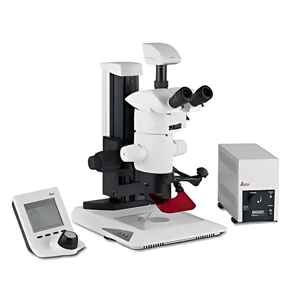
Leica MZ10F Modular Stereo Microscope
| Brand | Leica |
|---|---|
| Origin | Germany |
| Model | MZ10F |
| Magnification Range | 8×–80× |
| Resolution | 375 lp/mm |
| Illumination Architecture | TripleBeam™ Fluorescence-Specific Optical Path |
| Filter Changer | Fluo III Fast Filter Turret (4-position) |
| Modularity | Fully Modular Platform with Interchangeable Objectives, Illuminators, Cameras, and Ergonomic Stands |
Overview
The Leica MZ10F is a high-performance modular stereo microscope engineered for demanding fluorescence and brightfield applications in life science research, developmental biology, and genetic expression analysis. Built upon Leica’s proprietary TripleBeam™ optical architecture, the MZ10F separates the fluorescence excitation path from the observation and transmitted light paths—eliminating crosstalk, minimizing background noise, and maximizing signal-to-noise ratio in low-light fluorescent imaging. This dedicated beam routing ensures consistent illumination homogeneity and preserves fluorophore integrity during prolonged acquisition. The system delivers a continuous zoom magnification range of 8× to 80× (with 10× eyepieces), supported by an optical resolution of 375 line pairs per millimeter—enabling crisp visualization of subcellular structures, fine tissue morphology, and dynamic developmental processes in live or fixed specimens. Designed and manufactured in Wetzlar, Germany, the MZ10F adheres to stringent DIN ISO 9001 quality control standards and integrates seamlessly into GLP-compliant laboratory workflows.
Key Features
- TripleBeam™ Optical Design: Three physically independent light paths—one for fluorescence excitation, one for observation, and one for transmitted illumination—ensure optimal contrast, minimal photobleaching, and artifact-free multichannel imaging.
- Fluo III Fast Filter Turret: A motorized 4-position filter changer enabling sub-second switching between standard or user-defined fluorescence filter sets (e.g., DAPI/FITC/TRITC/Cy5), supporting rapid multi-label acquisition without manual intervention.
- High-Resolution Zoom Optics: Precision-ground apochromatic zoom optics provide 10:1 magnification ratio (8×–80×) with constant focus throughout zooming and edge-to-edge flatness across the field of view.
- Modular Hardware Ecosystem: Compatible with Leica’s full suite of accessories—including incident and transmitted LED illuminators (e.g., TL4000 LED, KL2500 LCD), ergonomic stands (e.g., S6, S8), digital cameras (DFC series), and motorized Z-drive units—allowing configuration tailored to dissection, documentation, or automated imaging tasks.
- Ergonomic & Service-Oriented Design: Adjustable interpupillary distance, diopter compensation, and tilting binocular tube support extended operator comfort; all optical components are factory-aligned and serviceable under Leica’s global calibration network.
Sample Compatibility & Compliance
The MZ10F accommodates a broad range of biological and material samples—from whole-mount zebrafish embryos and Drosophila larvae to plant meristems, histological sections, and microelectronic components. Its large working distance (up to 110 mm with optional objectives) enables manipulation under the objective using micromanipulators or patch-clamp rigs. The system meets IEC 61000-6-3 (EMC emissions) and IEC 61000-6-2 (immunity) standards. When paired with Leica Application Suite X (LAS X) software, it supports audit trails, electronic signatures, and data integrity features compliant with FDA 21 CFR Part 11 and EU Annex 11 requirements for regulated environments.
Software & Data Management
Control and image acquisition are managed via Leica Application Suite X (LAS X), a validated platform offering real-time Z-stack acquisition, multi-channel fluorescence registration, measurement tools (length, area, angle), and batch processing workflows. LAS X supports TIFF, JPEG2000, and Leica’s native .lei format with embedded metadata (objective ID, filter set, exposure time, stage coordinates). Raw data export is compatible with third-party analysis platforms including ImageJ/Fiji, Imaris, and MATLAB. All instrument settings and acquisition logs are timestamped and stored with user attribution—enabling full traceability in QA/QC and publication-ready documentation.
Applications
- Developmental biology: Time-lapse imaging of embryogenesis, organogenesis, and neural crest migration in model organisms (zebrafish, Xenopus, chick).
- Gene expression analysis: In situ hybridization (ISH) and immunofluorescence (IF) on whole-mount or sectioned tissues.
- Microdissection & electrophysiology: High-contrast visualization during laser capture microdissection (LCM) or patch-clamp electrode positioning.
- Quality control in biomanufacturing: Visual inspection of cell culture flasks, bioreactor harvests, and final product vials per USP and guidelines.
- Materials science: Surface topography assessment of MEMS devices, solder joints, and thin-film coatings under variable lighting conditions.
FAQ
Does the MZ10F support motorized focus or stage control?
Yes—when equipped with the optional Motorized Z-Drive (MZD) and/or Motorized XY Stage (MXY), the system enables programmable focus stacking and tiled scanning, fully controllable via LAS X.
Can I use third-party fluorescence filter sets with the Fluo III turret?
Yes—the Fluo III accepts standard 25 mm diameter filter cubes; custom sets from Chroma, Semrock, or Omega can be installed following Leica’s mechanical alignment protocol.
Is the MZ10F compatible with live-cell imaging chambers?
Yes—its long working distance and flexible illumination options (including ring LED and coaxial fiber-optic light guides) support integration with heated, CO₂-regulated, and humidity-controlled environmental chambers.
What is the warranty and service coverage for the MZ10F?
Leica provides a standard 24-month limited warranty covering parts and labor; extended service plans with preventive maintenance and priority response are available through authorized Leica Service Centers worldwide.
How does TripleBeam™ differ from conventional epi-fluorescence stereo microscopes?
Unlike shared-path systems where excitation light leaks into the detection path, TripleBeam™ uses discrete optical channels—reducing autofluorescence, improving contrast by >40% (measured at 488 nm excitation), and extending fluorophore lifetime during repeated imaging sessions.


