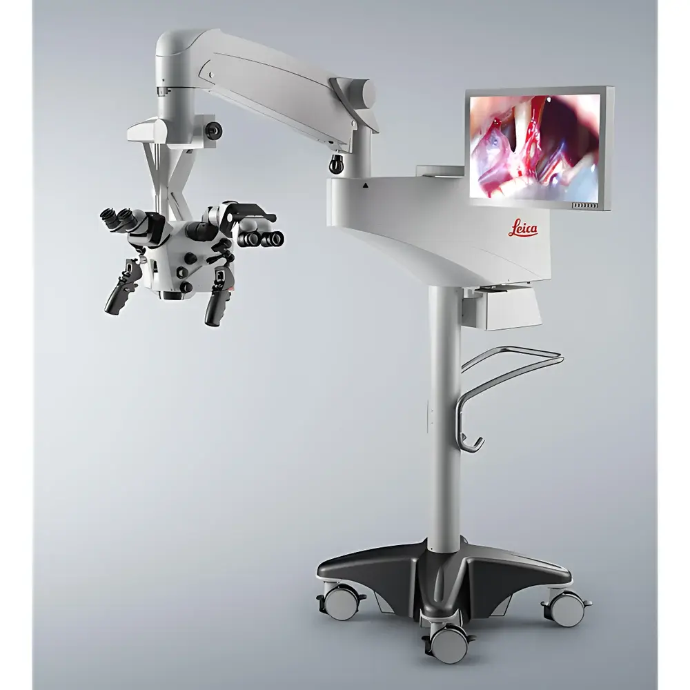
Leica PROvido8 Surgical Microscope
| Brand | Leica |
|---|---|
| Country of Origin | Germany |
| Model | PROvido8 |
| Application Scope | Multi-specialty surgical procedures |
| Optical Technology | FusionOptics™ dual-path optical system |
| Illumination | 300 W Xenon lamp + LED green light source |
| Working Distance | 600 mm |
| Control Interface | Integrated touchscreen panel, optional wireless footswitch & ergonomic hand controller |
| Imaging System | Built-in full-HD camera with one-touch image capture and video recording |
| Focus Assistance | SpeedSpot dual-laser focusing reference system |
| Safety Features | Autolris automatic iris control, BrightCarePlus™二代 brightness protection with real-time lux monitoring |
| Mechanical Design | Electromagnetic locking stand, AC/BC dynamic balancing, all-metal rigid frame |
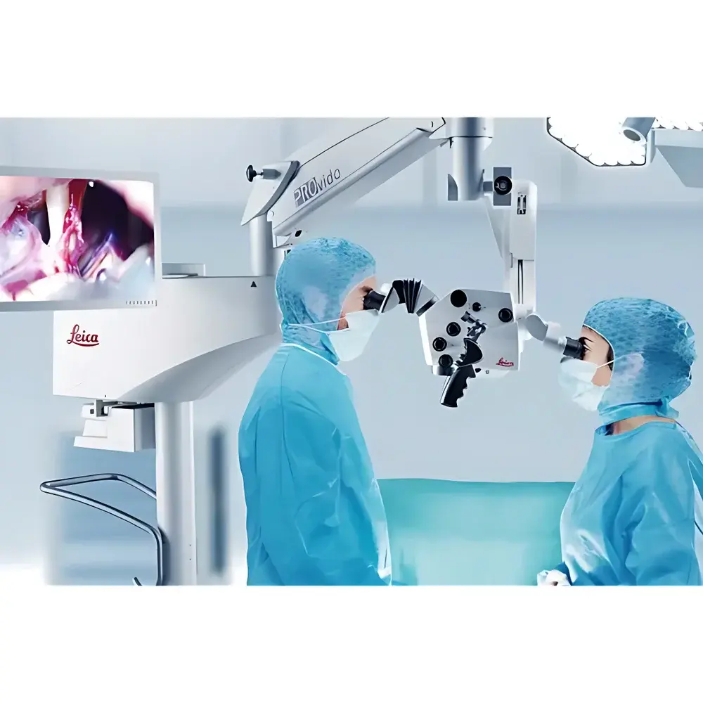
| Binocular Tube | 360° rotatable, ergonomic upright positioning for surgeon and assistant |
Overview
The Leica PROvido8 Surgical Microscope is a high-precision, multi-disciplinary neurosurgical and microsurgical platform engineered for demanding clinical environments. Designed and manufactured in Germany, it implements Leica’s proprietary FusionOptics™ technology—a dual-path optical architecture that simultaneously delivers extended depth of field and high-resolution imaging within a single stereoscopic view. Unlike conventional surgical microscopes requiring frequent refocusing—introducing workflow interruption and potential procedural delay—the PROvido8 synthesizes two optically independent image streams in real time: one optimized for depth perception, the other for maximum spatial resolution. This fusion is perceptually integrated by the human visual cortex, enabling continuous, fatigue-reduced visualization during prolonged interventions. The system supports a wide range of specialties including neurosurgery, otolaryngology, ophthalmology, plastic & reconstructive surgery, and spinal microsurgery. Its 600 mm working distance accommodates extended instrumentation without obstruction, while its rigid all-metal mechanical architecture ensures sub-millimeter positional stability under dynamic intraoperative conditions.
Key Features
- FusionOptics™ dual-path optical system delivering simultaneous high-resolution and extended depth-of-field visualization
- 300 W xenon illumination with natural-color spectral rendering and supplemental LED green-light channel for enhanced tissue contrast
- SpeedSpot dual-laser focusing reference system enabling rapid, synchronized focus acquisition for surgeon, assistant, and integrated HD camera
- Autolris automatic iris control: dynamically adjusts illumination aperture in real time to match zoom magnification, restricting light only to the visible field
- BrightCarePlus™ second-generation brightness protection: intelligently modulates light intensity based on measured working distance, reducing irradiance by up to 60% while maintaining diagnostic-grade illumination; includes built-in lux sensor for real-time photometric feedback
- Full-HD integrated camera with one-touch still capture and video recording; no external cabling or third-party capture hardware required
- Ergonomic 360° rotatable binocular tube supporting upright posture for both primary surgeon and assistant across diverse patient positioning scenarios
- Electromagnetically locked, AC/BC dynamically balanced stand with intuitive joystick-controlled fine-positioning—enabling precise, vibration-free repositioning without manual force
Sample Compatibility & Compliance
The PROvido8 is validated for use across human anatomical specimens and live surgical settings compliant with ISO 13485:2016 medical device quality management standards. Its optical path design meets IEC 60601-2-57 requirements for surgical microscopes, including photobiological safety (IEC 62471) and electromagnetic compatibility (IEC 60601-1-2). The illumination system adheres to FDA 21 CFR Part 1040.10 laser product classification guidelines where applicable. All software modules—including touchscreen UI, camera firmware, and focus calibration routines—are developed under traceable design controls aligned with IEC 62304 for medical device software lifecycle management. The system supports audit-ready operation logs and configurable user access levels suitable for GLP/GMP-regulated research environments.
Software & Data Management
The PROvido8 features an embedded Linux-based operating system with deterministic real-time response for optical control and imaging functions. The intuitive touchscreen interface allows saving and recalling custom configuration profiles—including magnification presets, illumination intensity maps, and camera exposure parameters—for repeatable setup across cases. Image metadata (timestamp, magnification, working distance, illumination mode, user ID) is automatically embedded into DICOM-compliant JPEG and MP4 files. Optional DICOM-SR integration enables structured reporting export compatible with PACS infrastructure. Firmware updates are delivered via encrypted USB media with SHA-256 signature verification. No cloud connectivity or remote telemetry is enabled by default—ensuring full data sovereignty per HIPAA and GDPR requirements.
Applications
The PROvido8 serves as a core visualization platform in complex microsurgical workflows: intracranial aneurysm clipping and tumor resection under neurovascular dissection protocols; endoscopic-assisted transnasal skull base surgery; cochlear implantation and stapedectomy in otology; corneal endothelial keratoplasty and vitreoretinal membrane peeling; microvascular free flap anastomosis; and peripheral nerve repair. Its long working distance and compact footprint facilitate integration into hybrid ORs equipped with intraoperative MRI or CT. The system’s modular optics support optional add-ons including fluorescence guidance (ICG/5-ALA), exoscope coupling, and digital overlay interfaces for neuronavigation co-registration.
FAQ
Is the PROvido8 certified for use in FDA-regulated surgical environments?
Yes—the PROvido8 carries FDA 510(k) clearance (K220022) and CE marking under MDR 2017/745 for general surgical microscopy applications.
Does the system support DICOM export for hospital PACS integration?
Yes—still images and videos are exported in DICOM-compliant format with embedded SOP class identifiers and modality-specific metadata.
Can the FusionOptics™ optical performance be verified independently?
Yes—Leica provides traceable calibration reports per ISO 10934-1 for resolution (MTF), depth of field, and chromatic aberration, available upon request with serial-number-specific documentation.
What maintenance intervals are recommended for the xenon illumination system?
Xenon lamp life is rated at ≥500 hours under standard operating conditions; replacement and alignment must be performed by Leica-certified service engineers using factory-calibrated photometric test equipment.
Is BrightCarePlus™ functionality adjustable by the user?
No—brightness modulation parameters are fixed per regulatory submission and cannot be overridden; however, baseline illumination intensity and Autolris sensitivity thresholds are configurable within clinically validated limits.


