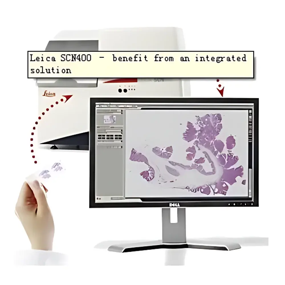
Leica SCN400 Slide Scanning System
| Brand | Leica |
|---|---|
| Origin | Germany |
| Model | SCN400 |
| Automation | Integrated SL801 384-slide autoloader |
| Max Magnification | 40× |
| Avg. Scan Time (20×) | 100 s per slide |
| Focusing Technology | Patented Dynamic Focus Tracking |
| Software Platform | Leica SCN400 Client |
Overview
The Leica SCN400 Slide Scanning System is a high-throughput, research-grade whole-slide imaging (WSI) platform engineered for precision digital pathology and histology laboratories. Based on automated brightfield microscopy with motorized XYZ-stage control and multi-objective turret configuration, the system captures high-fidelity, focus-stable digital replicas of glass-mounted tissue sections at magnifications up to 40×. Its core innovation lies in the patented Dynamic Focus Tracking technology—a real-time optical feedback loop that continuously adjusts objective position during scanning to compensate for microscopic topographic variations across stained slides (e.g., uneven mounting medium, section thickness gradients, or cover slip curvature). Unlike conventional pre-scan focus mapping or z-stack acquisition methods, this closed-loop approach eliminates time-intensive calibration steps while maintaining sub-micron axial stability—enabling consistent optical sectioning without user intervention or re-scans.
Key Features
- Dynamic Focus Tracking: Proprietary hardware-accelerated autofocus algorithm operating at ≥50 Hz sampling rate, ensuring continuous focal plane maintenance across heterogeneous slide surfaces.
- SL801 Autoloader Integration: Robust 384-position robotic slide loader compliant with ANSI/SLAS microplate footprint standards; supports unattended batch scanning with barcode-based slide identification and priority queuing.
- Multi-Protocol Concurrent Execution: Software-controlled parallel processing of up to eight independent scanning protocols—including variable magnification, channel selection (brightfield only), region-of-interest (ROI) definition, and compression settings—within a single run.
- Optical Architecture: Plan-apochromat objectives (20× and 40×, NA ≥0.75), LED illumination with color temperature stabilization (5700 K ±50 K), and 16-bit monochrome CMOS sensor (6.5 µm pixel pitch) optimized for IHC and H&E contrast fidelity.
- Thermal & Mechanical Stability: Precision-machined aluminum chassis with active thermal management; positional repeatability <±0.2 µm over 8-hour operation under ambient lab conditions (20–25°C).
Sample Compatibility & Compliance
The SCN400 accepts standard 1″ × 3″ (25 × 75 mm) glass slides with thicknesses from 0.9 to 1.2 mm, including charged, poly-L-lysine-coated, and silanized variants. It accommodates common histological preparations: formalin-fixed paraffin-embedded (FFPE) sections (3–5 µm), frozen sections, cytology smears, and hematopathology blood films. The system complies with ISO 13485:2016 for medical device quality management and supports audit-ready operation under GLP and CAP-accredited workflows. Digital output formats include DICOM-SR (Supplement 145), TIFF (uncompressed and LZW-compressed), and JPEG2000—fully compatible with PACS integration and FDA 21 CFR Part 11–enabled LIMS environments when deployed with validated Leica Biosystems’ enterprise software modules.
Software & Data Management
The Leica SCN400 Client is a Windows-based application built on Microsoft .NET Framework 4.8, featuring role-based access control (RBAC), encrypted local database storage (SQL Server Express), and configurable metadata tagging per slide (e.g., patient ID, staining protocol, pathologist annotation). It generates standardized QC reports—including focus confidence metrics, illumination uniformity maps, and slide-level artifact detection logs—for internal validation and regulatory review. Raw image data is stored in vendor-neutral hierarchical data format (HDF5) with embedded EXIF-like metadata, enabling interoperability with third-party AI training pipelines (e.g., QuPath, HALO, Visiopharm). Optional Leica eSlide Manager provides centralized deployment, remote monitoring, and DICOMweb™ RESTful API access for cloud-based image retrieval.
Applications
The SCN400 serves as a foundational imaging engine in academic pathology departments, contract research organizations (CROs), and pharmaceutical translational labs. Primary use cases include: retrospective tissue biomarker quantification in clinical trial cohorts; digital archiving of rare disease repositories; telepathology consultation networks requiring ultra-high-resolution static imaging; computational pathology algorithm development (e.g., tumor segmentation, mitotic count automation); and routine QC of immunohistochemistry assay performance across multi-site studies. Its deterministic scan timing and focus resilience make it especially suited for longitudinal studies where inter-scan comparability across months or years must be preserved without recalibration.
FAQ
Does the SCN400 support fluorescence scanning?
No—the SCN400 is a brightfield-only platform. Fluorescence WSI requires the Leica GT450 or Aperio AT2 systems.
Can scanned images be exported directly to a PACS?
Yes, via DICOM-SR export with configurable SOP Class UIDs and modality worklist integration using standard HL7 v2.5.1 interfaces.
Is dynamic focus tracking available for all magnifications?
Yes—it operates continuously at 20× and 40× objectives; 1.25× and 2.5× overview modes use fixed-focus interpolation.
What is the maximum supported slide throughput per 24-hour period?
Under optimal conditions (20×, full-slide scan, SL801 loaded), the system achieves ~300–330 slides/day with <5% re-scan rate due to focus failure.
Does Leica provide IQ/OQ/PQ documentation for GxP environments?
Yes—validated installation qualification (IQ), operational qualification (OQ), and performance qualification (PQ) protocols are available upon request and align with ASTM E2500-13 and USP guidelines.


