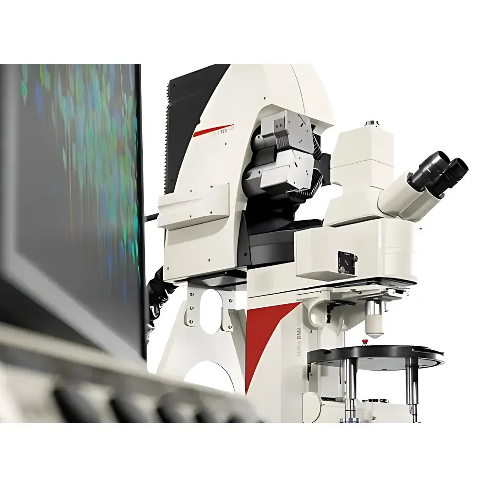
Leica TCS SP8 MP Multiphoton Confocal Microscope
| Brand | Leica |
|---|---|
| Origin | Germany |
| Model | TCS SP8 MP |
| Instrument Type | Point-Scanning Multiphoton Confocal Microscope |
| Excitation Range | 680–1300 nm |
| Maximum IR Wavelength | 1300 nm |
| Resonant Scanner Speed | 8 kHz / 12 kHz |
| Field of View (FOV) | up to 22 mm |
| Detection | Leica HyD NDD & HyD RLD non-descanned detectors |
| Objective Correction | motCORR electrically adjustable correction collar |
| Specialized Objectives | HC IRAPO L 25×/1.00 W motCORR, HC FLUOTAR L 25×/1.00 IMM (ne=1.457) motCORR VISIR, APO HCX L20×/0.95 IMM for BABB immersion (ne=1.559) |
| Software Platform | LAS X with integrated multiphoton control, FLIM wizard, and STED/FLIM/SMD module compatibility |
| Compliance | Designed for GLP/GMP-aligned workflows |
Overview
The Leica TCS SP8 MP is a high-performance, modular multiphoton confocal microscope engineered for deep-tissue, live-specimen imaging with minimal phototoxicity and maximal signal fidelity. Unlike conventional visible-light confocal systems—where scattering and out-of-focus excitation limit penetration depth and compromise viability—the TCS SP8 MP leverages near-infrared (NIR) and extended infrared (IR) multiphoton excitation (680–1300 nm) to achieve superior optical sectioning in highly scattering biological specimens. Its core principle relies on simultaneous absorption of two or more long-wavelength photons only at the focal plane, eliminating axial background and enabling intrinsic 3D resolution without pinhole-based optical sectioning. This nonlinear excitation mechanism significantly reduces photobleaching and photodamage outside the focal volume, preserving cellular physiology during prolonged time-lapse experiments. The system integrates a robust point-scanning architecture with dual resonant galvanometric scanners, motorized optical components, and a fully synchronized laser control interface—making it suitable for quantitative neuroscience, developmental biology, organ-level CLARITY/BABB imaging, and dynamic subcellular process analysis.
Key Features
- Integrated multiphoton excitation platform supporting tunable femtosecond lasers (e.g., Insight DeepSee+, OPO MPX) with seamless wavelength selection from 680 nm to 1300 nm via LAS X software
- Leica HyD NDD and HyD RLD non-descanned detectors delivering photon-counting sensitivity, high quantum efficiency (>45% at 800 nm), and sub-nanosecond timing resolution for FLIM applications
- motCORR electrically actuated correction collars on specialized objectives (e.g., HC IRAPO L 25×/1.00 W, HC FLUOTAR L 25×/1.00 IMM VISIR) for real-time compensation of refractive index mismatch across depth, temperature, and mounting medium variations
- High-speed resonant scanning at 8 kHz and 12 kHz, enabling frame rates up to 428 fps (512 × 16 px) or 40 fps (512 × 512 px), coupled with SuperZ GalvoFlow for accelerated 4D stack acquisition
- 22 mm maximum field-of-view scanning capability using Leica’s patented x2y mirror design—largest FOV among commercial point-scanning multiphoton systems
- Quad detection module supporting up to four parallel HyD channels for simultaneous multicolor multiphoton imaging, SHG/THG detection, and electrophysiology-correlated acquisition
Sample Compatibility & Compliance
The TCS SP8 MP is validated for imaging diverse specimen types—from single living cells and acute brain slices to cleared whole organs (up to 6 mm depth) processed with CLARITY, BABB, or similar hydrophilic/hydrophobic clearing protocols. Its dedicated objective portfolio includes water-immersion (ne ≈ 1.33), glycerol-immersion (ne = 1.45), and BABB-immersion (ne = 1.559) optics, each optimized for chromatic correction across both single-photon (VIS) and multiphoton (NIR–IR) spectral bands. The system supports ISO/IEC 17025-aligned calibration procedures and is compatible with laboratory quality management frameworks requiring traceable instrument qualification. When configured with LAS X software in validated mode—including user role management, electronic signatures, and full audit trail logging—it meets documentation requirements for regulated environments governed by FDA 21 CFR Part 11, EU Annex 11, and GLP/GMP standards.
Software & Data Management
LAS X serves as the unified control and analysis environment for the TCS SP8 MP, offering intuitive, workflow-driven wizards for multiphoton setup, FLIM acquisition, STED alignment, and spectral unmixing. Its modular architecture allows integration of third-party libraries (e.g., PicoQuant’s SymPhoTime for advanced FLIM-FRET analysis) while maintaining native synchronization with hardware timing engines. All acquisition parameters—including laser power, dwell time, z-step size, detector gain, and motCORR position—are stored within the proprietary .lif metadata format, ensuring full experimental reproducibility. Raw data export supports TIFF, HDF5, and OME-TIFF standards for interoperability with ImageJ/Fiji, Python-based analysis pipelines (e.g., Napari, scikit-image), and cloud-based HPC platforms. Automated batch processing, ROI-based quantification, and multi-dimensional histogram analysis are embedded natively, reducing post-acquisition overhead without reliance on external scripting.
Applications
The TCS SP8 MP addresses demanding use cases where depth, speed, and molecular specificity converge. In neurobiology, it enables longitudinal calcium dynamics mapping in intact cortical slabs or whole-brain cleared samples, revealing circuit-level connectivity beyond the limits of serial sectioning. For developmental studies, its combination of low phototoxicity and high NA immersion optics facilitates long-term imaging of embryonic organogenesis in live zebrafish or mouse embryos. In immunology and oncology, simultaneous multichannel detection supports tracking of immune cell infiltration, tumor microenvironment remodeling, and vascular permeability changes in orthotopic models. Its SHG/THG capability further extends label-free structural imaging—particularly valuable for collagen fiber orientation analysis in fibrotic tissues or myosin organization in muscle biopsies. Additionally, the platform’s FLIM-ready architecture supports quantitative FRET biosensor readouts, metabolic redox state assessment via NAD(P)H lifetime, and protein conformational dynamics in situ.
FAQ
What distinguishes multiphoton excitation from standard confocal imaging?
Multiphoton excitation uses longer-wavelength, lower-energy photons that only generate fluorescence when two or more photons are absorbed simultaneously at the focal point—eliminating out-of-focus excitation and enabling deeper penetration with reduced photodamage.
Can the TCS SP8 MP be upgraded to support super-resolution techniques?
Yes. The modular SP8 platform supports seamless integration of STED 3X, white light laser (WLL), fluorescence lifetime imaging (FLIM), and stimulated emission depletion modules—all controlled through the same LAS X interface.
Is motCORR correction required for all sample types?
It is essential for thick, heterogeneous, or optically cleared specimens where refractive index gradients cause spherical aberration; however, it may be disabled for thin, homogeneous monolayers imaged with matched immersion media.
How does the system handle spectral crosstalk in multicolor multiphoton experiments?
Through precise laser line selection, sequential or interleaved excitation schemes, and linear unmixing algorithms applied to spectrally resolved HyD data—enabling clean separation of fluorophores with overlapping emission profiles.
What validation documentation is available for regulated research environments?
Leica provides IQ/OQ documentation templates, laser power calibration certificates, and LAS X 3.7+ validation support packages—including risk assessments, UAT scripts, and 21 CFR Part 11 compliance checklists—for GxP-aligned deployments.


