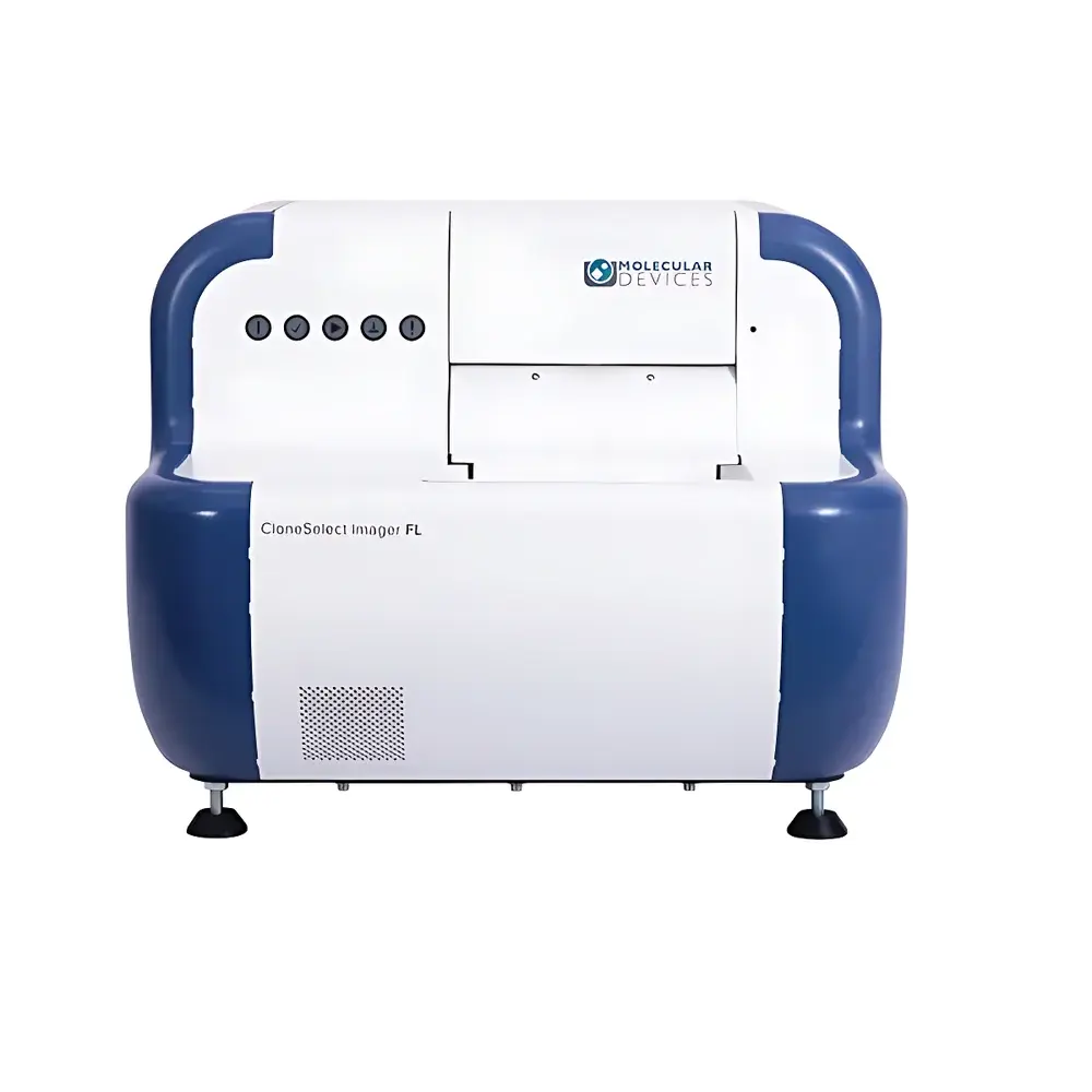
Molecular Devices CloneSelect Imager FL Cell Growth and Clonality Analysis System
| Brand | Molecular Devices |
|---|---|
| Origin | Imported |
| Manufacturer Type | Original Equipment Manufacturer (OEM) |
| Model | CloneSelect Imager FL |
| Pricing | Available upon Request |
Overview
The Molecular Devices CloneSelect Imager FL is a fully automated, non-invasive cell growth and clonality analysis system engineered for high-content, label-free imaging of adherent and suspension cultures in standard microplates. Leveraging dual-mode optical architecture—simultaneous brightfield and multi-channel fluorescence imaging—the system captures kinetic cellular data with temporal precision from Day 0 onward. Its core measurement principle relies on quantitative pixel-based image analysis to compute confluence, cell count, clone area, and growth kinetics without dyes, labels, or endpoint assays. Designed specifically for therapeutic monoclonal antibody (mAb) development and cell line development (CLD), the Imager FL supports regulatory-compliant workflows aligned with ICH Q5D, USP , and FDA guidance on clonality assurance for IND submissions (21 CFR Part 312). It delivers objective, auditable evidence of monoclonality by detecting and validating the “locus of growth” from single-cell seeding events—a critical requirement for biologics manufacturing under current Good Manufacturing Practice (cGMP) frameworks.
Key Features
- Simultaneous brightfield and up to four-channel fluorescence imaging (e.g., GFP, RFP, DAPI, Cy5) with motorized filter wheels and LED excitation sources optimized for low phototoxicity and long-term kinetic studies
- Automated plate scanning at user-defined intervals (minutes to days), supporting 6–384-well formats with focus stabilization across well edges and variable well depths
- Real-time confluence calculation per well using adaptive thresholding algorithms validated against manual hemocytometer counts (R² > 0.98)
- Single-cell detection and tracking from Day 0 via high-resolution (≥1.2 µm/pixel) imaging and machine learning–enhanced segmentation
- Integrated “Loci of Growth” algorithm that identifies wells originating from a single viable cell, flagging ambiguous or multi-locus events for review
- Regulatory-ready audit trail with timestamped metadata, user actions, instrument settings, and raw image provenance—fully compliant with 21 CFR Part 11 requirements
Sample Compatibility & Compliance
The CloneSelect Imager FL accommodates diverse mammalian cell types including CHO-K1, CHO-S, HEK293, hybridoma, iPSCs, and primary T cells—both adherent and non-adherent (e.g., suspension-adapted lines settled via centrifugation or gravity sedimentation). It operates within standard biosafety level 2 (BSL-2) laboratory environments and integrates seamlessly into ISO 13485–certified QC labs and GLP/GMP-controlled cell banking suites. All image acquisition and analysis protocols are documented per internal SOPs and support traceability to ASTM E2500-22 (Standard Guide for Specification, Design, and Verification of Pharmaceutical and Biopharmaceutical Manufacturing Systems) and ICH Q5D (Quality of Biotechnological Products: Derivation and Characterization of Cell Substrates).
Software & Data Management
Powered by CloneSelect Software v5.x, the system provides a secure, role-based interface with configurable user permissions, electronic signatures, and full ALCOA+ (Attributable, Legible, Contemporaneous, Original, Accurate, Complete, Consistent, Enduring, Available) data handling. Raw images, processed metrics (confluence %, cell count, doubling time, clone area), and growth curves are stored in a relational database with automated backup and version control. Export options include CSV, TIFF, PNG, PDF, and Microsoft Word—enabling direct inclusion of validated reports in regulatory dossiers. The software supports batch processing, custom analysis templates, and API access for integration with LIMS (e.g., LabVantage, Thermo Fisher SampleManager) and ELN platforms.
Applications
- Clonality verification for master cell bank (MCB) generation and IND-enabling studies
- CRISPR/Cas9 and other gene-editing validation via co-expression of fluorescent reporters and functional markers
- Cell line stability assessment through longitudinal confluence and morphology tracking over ≥30 passages
- Cytotoxicity profiling using fluorescence-based viability indicators (e.g., Calcein AM/EthD-1) or label-free confluence decline kinetics
- Process parameter optimization (e.g., seeding density, feed strategy, matrix coating) during early-stage CLD
- Comparative growth analysis across clones to select high-producer candidates with optimal expansion kinetics
FAQ
Does the CloneSelect Imager FL require cell staining or labeling to quantify confluence?
No. It performs label-free confluence analysis using calibrated brightfield imaging and proprietary segmentation algorithms trained on morphologically diverse cell types.
Can the system validate monoclonality for FDA submission under 21 CFR Part 312?
Yes. The “Monoclonality Report” module generates audit-ready documentation—including timestamped Day 0 single-cell images, loci-of-growth annotations, and reviewer sign-offs—that satisfies FDA expectations for clonality evidence in IND applications.
Is the software compliant with 21 CFR Part 11 for electronic records and signatures?
Yes. CloneSelect Software includes built-in Part 11 controls: electronic signatures with identity verification, immutable audit trails, and system validation documentation available upon request.
What fluorescence channels are supported out of the box?
Standard configuration includes GFP (470/22 nm ex, 525/50 nm em), RFP/mCherry (560/25 nm ex, 630/60 nm em), DAPI (365/12 nm ex, 445/50 nm em), and Cy5 (625/30 nm ex, 700/75 nm em), with optional filter upgrades available.
How does the system handle edge effects or meniscus distortion in low-volume wells?
The autofocus routine employs multi-point z-stack acquisition per well, combined with meniscus-aware ROI definition and background correction algorithms to maintain accuracy in 384- and 1536-well plates.


