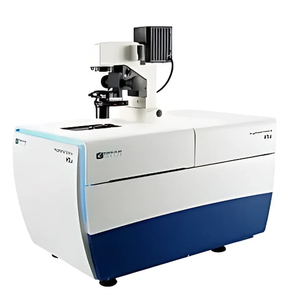
Molecular Devices ImageXpress Confocal HT.ai Intelligent High-Content Confocal Imaging and Analysis System
| Brand | Molecular Devices |
|---|---|
| Origin | USA |
| Model | ImageXpress Confocal HT.ai |
| Environmental Control | 30–40 °C ± 0.5 °C |
| Laser Channels | 7-color solid-state lasers |
| Imaging Channels | 8 (DAPI, CFP, FITC, YFP, TRITC, Texas Red, Cy5, Cy7) |
| Objective Compatibility | Up to 25 objectives including oil (NA 1.4), air (NA 0.05–0.95), and water immersion (NA 1.2) |
| Detection | sCMOS camera |
| Confocal Technology | AgileOptix™ spinning-disk confocal |
| Software Suite | MetaXpress® + IN Carta™ |
| Compliance | Supports GLP/GMP workflows, FDA 21 CFR Part 11 audit trail (via optional configuration), ISO/IEC 17025 traceability-ready |
Overview
The Molecular Devices ImageXpress Confocal HT.ai is an intelligent, high-content confocal imaging and analysis system engineered for quantitative, multi-parametric phenotypic profiling of complex 3D biological models—including organoids, spheroids, thick tissue sections, zebrafish embryos, and C. elegans—in physiologically relevant microenvironments. It operates on the principle of spinning-disk confocal microscopy, leveraging AgileOptix™ optical architecture to eliminate out-of-focus light while maintaining high-speed acquisition and deep optical sectioning capability. Unlike widefield systems, this platform delivers true optical sectioning with enhanced axial resolution (typically <1 µm), enabling precise Z-stack reconstruction and volumetric quantification across heterogeneous samples up to 500 µm in thickness. Integrated environmental control—precisely regulated at 30–40 °C ± 0.5 °C, with optional CO2 and humidity modules—ensures long-term viability during kinetic assays spanning minutes to days. The system is purpose-built for drug discovery, functional genomics, neurobiology, and regenerative medicine applications where spatial fidelity, signal-to-noise ratio, and statistical robustness are non-negotiable.
Key Features
- AgileOptix™ spinning-disk confocal technology with interchangeable disk configurations and seven solid-state laser lines (405, 445, 488, 514, 561, 594, 640 nm), enabling flexible excitation matching for multiplexed fluorescent probes.
- Eight simultaneous detection channels supporting standard fluorophores: DAPI, CFP, FITC, YFP, TRITC, Texas Red, Cy5, and Cy7—optimized for subcellular to whole-tissue scale resolution.
- Water-immersion objective system (20×, 40×, 60×; NA 1.2) delivering up to 4× signal enhancement versus air objectives without compromising acquisition speed—critical for low-signal 3D samples embedded in Matrigel or collagen.
- sCMOS detector with >82% quantum efficiency, 16-bit dynamic range, and low read noise (<1.2 e−) ensures high-fidelity photon capture across wide intensity distributions.
- QuickID™ intelligent targeting workflow: low-magnification scouting followed by automated high-resolution, multi-Z, multi-channel reimaging of rare events or region-of-interest (ROI) within microplates—enabling unbiased, scalable screening of heterogeneous populations.
- Full motorized hardware integration: autofocus (laser-based + image-based), stage positioning, filter wheels, environmental chamber, and illumination—all synchronized via a deterministic real-time control engine.
Sample Compatibility & Compliance
The ImageXpress Confocal HT.ai accommodates diverse specimen formats—from 6- to 384-well plates (including U-bottom spheroid plates), chamber slides, and custom substrates—without mechanical reconfiguration. Its modular optical path supports oil (NA 1.4), air (0.05–0.95 NA), and water immersion (NA 1.2) objectives, permitting optimal resolution across sample thicknesses and refractive indices. For regulatory-compliant environments, the system supports audit-trail-enabled operation under FDA 21 CFR Part 11 when paired with validated MetaXpress® software configurations. Instrument qualification documentation (IQ/OQ/PQ) templates align with ISO/IEC 17025 and GLP requirements. All environmental parameters—including temperature, CO2, and humidity—are logged with timestamped metadata and exportable in CSV or HDF5 format for traceability.
Software & Data Management
MetaXpress® software provides unified control over acquisition, analysis, and data management. Its modular architecture includes pre-validated analysis modules for cell counting, nuclear morphology, organelle translocation, neurite outgrowth, and mitotic staging—each configurable via drag-and-drop workflow editors. Adaptive Background Correction™ dynamically adjusts thresholding based on local fluorescence intensity, improving segmentation accuracy in heterogeneous fields. For 3D analysis, the software computes volumetric metrics (e.g., object volume, surface area, XYZ centroid position, inter-object distance, texture heterogeneity) and exports per-cell or per-field summary statistics in standardized tabular formats. IN Carta™ complements MetaXpress® with machine learning–driven phenotypic classification: users define training sets via intuitive ROI annotation; the software trains convolutional neural networks (CNNs) to classify morphological states without manual feature engineering. Batch processing, parameter versioning, and REST API integration enable pipeline automation and LIMS connectivity.
Applications
This system serves as a central platform for quantitative phenotypic screening in academic, pharmaceutical, and contract research settings. Validated use cases include: high-content toxicity assessment in iPSC-derived cardiomyocytes; longitudinal tracking of organoid growth and lumen formation; compound-induced changes in mitochondrial network morphology; spatial mapping of immune cell infiltration in tumor spheroids; and functional readouts from CRISPR-edited neuronal cultures. Its ability to resolve subcellular structures (e.g., ER tubules, actin stress fibers, nuclear lamina) within intact 3D models enables translationally relevant endpoints—such as barrier integrity, ciliary beating frequency, or synaptic puncta density—that correlate strongly with in vivo pharmacodynamics.
FAQ
What distinguishes AgileOptix™ confocal technology from traditional pinhole-based systems?
AgileOptix™ uses a dual-disk design with variable pinhole spacing and optimized microlens arrays to balance optical sectioning strength with photon throughput—achieving ~90% transmission efficiency while maintaining axial resolution comparable to point-scanning confocals.
Can the system perform label-free imaging?
Yes—integrated brightfield, phase contrast, and DIC modules support label-free morphological assessment; combined with autofluorescence channel subtraction, they enable multimodal phenotyping without exogenous dyes.
Is IN Carta™ compatible with third-party image formats?
IN Carta™ accepts OME-TIFF, ND2, CZI, and LSM files; it also supports direct import from MetaXpress® project archives with full metadata preservation.
How is calibration traceability maintained?
NIST-traceable stage calibration slides and fluorescence intensity standards (e.g., Chroma QF100 series) are supported via built-in calibration wizards; all calibration events are time-stamped and stored in the audit log.
What level of IT infrastructure is required for deployment?
A dedicated Windows Server 2019/2022 host with ≥64 GB RAM, ≥2 TB SSD storage, and 10 GbE networking is recommended for concurrent acquisition and analysis of large 3D datasets; cloud backup and remote access are supported via secure HTTPS tunneling.


