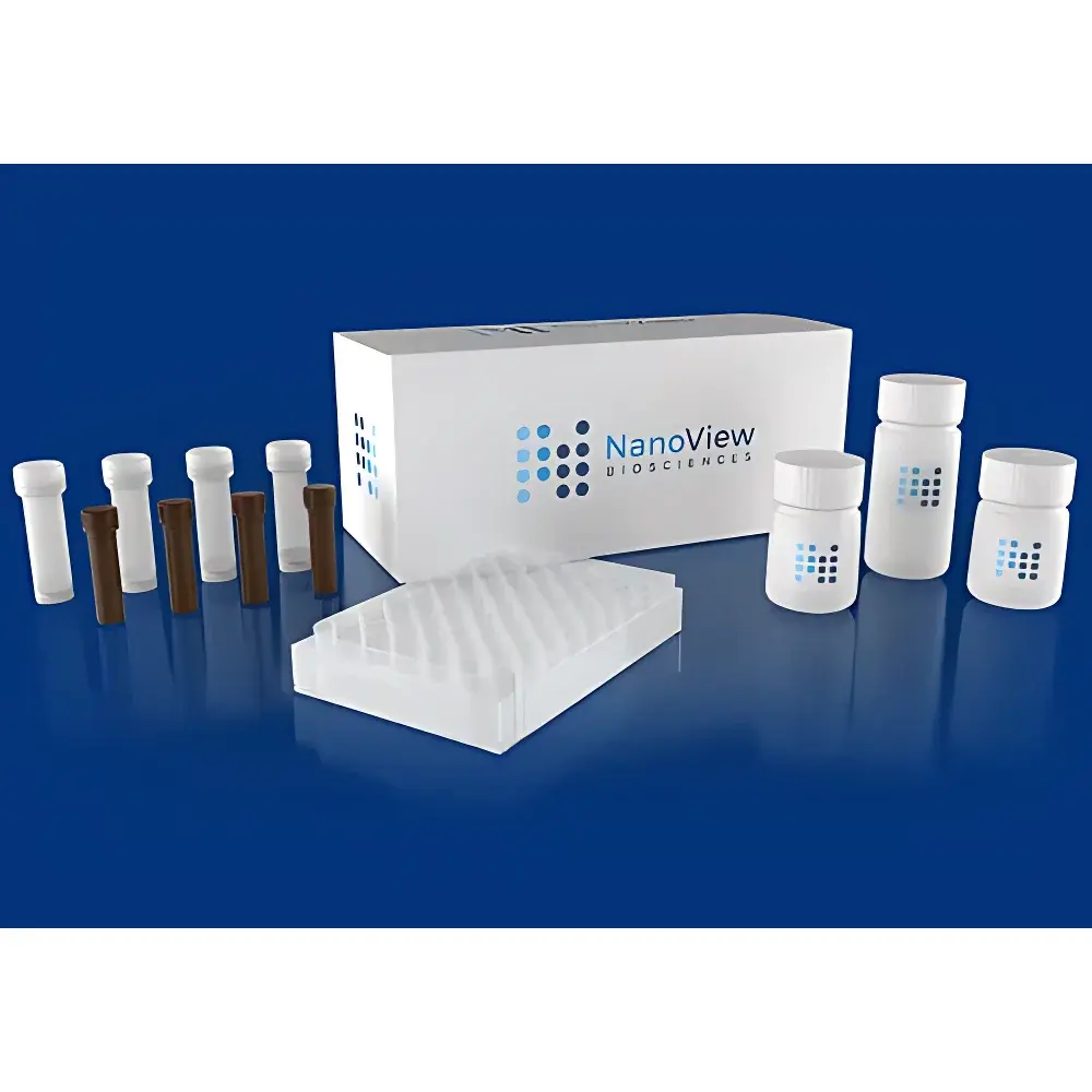
NanoView Biosciences ExoView Comprehensive Exosome Characterization Kit
| Brand | NanoView Biosciences |
|---|---|
| Origin | USA |
| Distribution Type | Authorized Distributor |
| Import Status | Imported |
| Catalog Variants | EV-TETRA-C, EV-TETRA-P, EV-TETRA-M2, EV-TETRA-C-CAR, EV-TETRA-P-CAR, EV-TC-FLEX, EV-TP-FLEX, EV-TC-FLEX-CAR, EV-TP-FLEX-CAR, EV-TM-FLEX, EV-TM-FLEX-CAR, EV-FLEX-2, EV-FLEX-2-CAR, EV-CTETRA-1/2/3, EV-CTETRA-1/2/3-CAR, EV-CUST-1–6, EV-CUST-1–6-CAR |
| Sample Volume | 35 µL |
| Replicates | Triplicate |
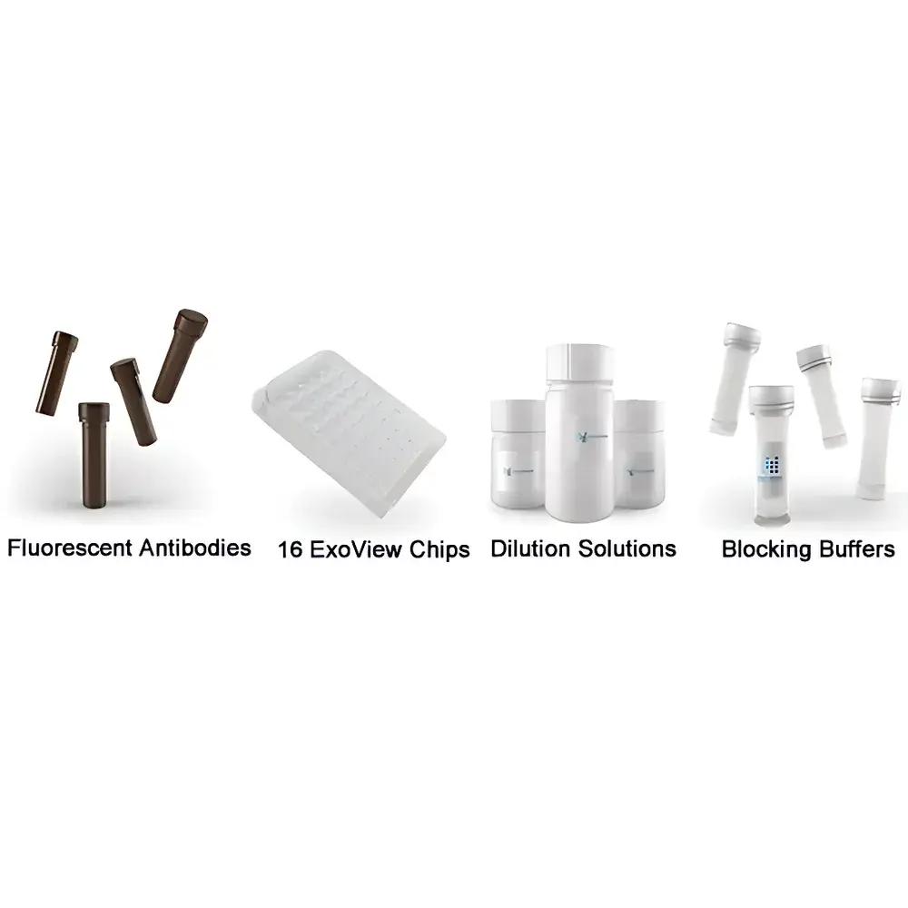
| Capture Antibodies | anti-CD9, anti-CD63, anti-CD81, IgG isotype control |
| Detection Fluorophores | CD9 (Blue), CD81 (Green), CD63 (Red) |
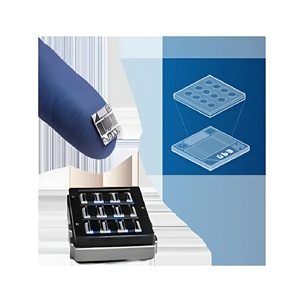
| Detection Principle | Single-Particle Interferometric Reflectance Imaging Sensing (SP-IRIS) |
| Minimum Detectable Size | ≥50 nm |
| Content Detection Capability | Yes (with membrane-permeabilizing reagent) |
| Compliance | Designed for GLP-compliant workflows |
Overview
The NanoView Biosciences ExoView Comprehensive Exosome Characterization Kit is a benchtop, chip-based assay system engineered for label-free, high-resolution single-particle analysis of extracellular vesicles (EVs) directly in complex biological matrices. It leverages Single-Particle Interferometric Reflectance Imaging Sensing (SP-IRIS) — a surface-sensitive optical detection method that quantifies individual exosomes immobilized on antibody-patterned silicon chips without centrifugation, ultracentrifugation, or size-exclusion chromatography. Unlike bulk measurement techniques (e.g., NTA, DLS, or ELISA), SP-IRIS enables simultaneous enumeration, sizing (≥50 nm resolution), phenotypic profiling (via multiplexed surface marker expression), and spatial co-localization analysis at the single-vesicle level. The platform is optimized for direct analysis of minimally processed samples including cell culture supernatants, human plasma, serum, urine, cerebrospinal fluid (CSF), and saliva — eliminating pre-analytical variability introduced by purification artifacts.
Key Features
- Multiplexed capture capability: Up to six distinct capture antibodies (e.g., anti-CD9, anti-CD63, anti-CD81, plus custom options) can be spatially arrayed on a single chip, enabling parallel enrichment of functionally or biologically defined exosome subpopulations.
- Single-vesicle resolution: SP-IRIS provides interferometric height measurements correlated with particle diameter, yielding quantitative size distribution histograms with sub-50 nm sensitivity — validated against TEM and cryo-EM reference standards.
- Co-localization quantification: Dual- or triple-color fluorescence imaging allows pixel-level correlation of surface markers (e.g., CD9Blue/CD63Red) on individual particles, supporting rigorous statistical inference of co-expression frequency and heterogeneity metrics.
- No purification required: Direct application of native biofluids preserves exosome integrity and avoids loss or aggregation associated with ultracentrifugation or polymer-based precipitation methods.
- Content-accessible profiling: Optional membrane-permeabilizing reagents enable intravesicular staining (e.g., Syntenin, miRNA probes), extending characterization beyond surface epitopes into cargo composition.
- Configurable assay formats: Modular kit architecture supports standardized (TETRA-series), flexible (FLEX-series), and fully customizable (CUST-series) configurations — accommodating both discovery and translational validation workflows.
Sample Compatibility & Compliance
The ExoView system demonstrates robust performance across diverse sample types without dilution or pre-filtration: cell culture supernatants (serum-free or FBS-depleted), EDTA-plasma, serum, urine, CSF, and salivary exudates. Each kit variant is validated for its designated matrix (e.g., EV-TETRA-P for plasma, EV-TETRA-M2 for murine models). All assays are compatible with Good Laboratory Practice (GLP) and current Good Manufacturing Practice (cGMP)-aligned environments. Data acquisition and reporting modules support electronic signatures, time-stamped audit trails, and secure user access controls compliant with FDA 21 CFR Part 11 when deployed on validated IT infrastructure. The platform aligns with MISEV2018 reporting guidelines and supports ISO 24705:2022 (Extracellular Vesicles — Requirements for Characterization) through traceable calibration and documented uncertainty budgets.
Software & Data Management
Data acquisition and analysis are performed using ExoView Analyzer Software (v3.2+), a Windows-based application featuring automated image segmentation, intensity thresholding, particle classification, and batch processing pipelines. Raw interferometric reflectance images and fluorescence channel stacks are stored in vendor-neutral TIFF format with embedded metadata (sample ID, date/time, operator, instrument settings). Export options include CSV (particle-by-particle attributes), PDF reports (summary statistics, co-localization heatmaps, size distributions), and HDF5 for integration with Python/R-based bioinformatics platforms. Audit logs record all user actions, parameter modifications, and report generations — essential for regulatory submissions and internal quality assurance reviews.
Applications
- Phenotypic mapping of disease-associated exosome subpopulations in oncology (e.g., PD-L1+/CD81+ tumor-derived EVs in melanoma plasma)
- Longitudinal monitoring of therapeutic EV release from MSC or iPSC-derived cell therapies
- Validation of EV isolation methods via orthogonal comparison to SEC, UC, or immunoaffinity capture
- Biomarker discovery in neurodegenerative disorders using CSF-derived exosomes enriched for L1CAM or NCAM
- Quality control of clinical-grade EV products under GMP conditions, including potency and consistency assessment
- Functional studies correlating surface proteome (e.g., integrin α6β4) with tissue tropism in preclinical models
FAQ
What sample volume is required per assay?
A single assay requires only 35 µL of undiluted or low-fold diluted biofluid — sufficient for triplicate analysis on one chip.
Can I use my own capture antibodies?
Yes. The FLEX and CUST series kits provide blank or pre-spotted chips compatible with user-defined antibodies, including species-specific or novel target binders.
How does SP-IRIS differ from conventional ELISA or flow cytometry?
SP-IRIS detects individual particles optically without hydrodynamic focusing or labeling-dependent amplification — avoiding signal saturation, coincidence errors, and antibody affinity bias inherent in ensemble or microbead-based approaches.
Is content detection compatible with RNA or protein cargo?
Yes. With appropriate permeabilization and probe design, the system supports fluorescent detection of intraluminal proteins (e.g., Syntenin, TSG101) and structured RNA motifs using hybridization probes.
Are calibration standards included?
Each kit includes NIST-traceable silica nanoparticle standards (50 nm, 100 nm, 200 nm) for routine system verification and size calibration.


