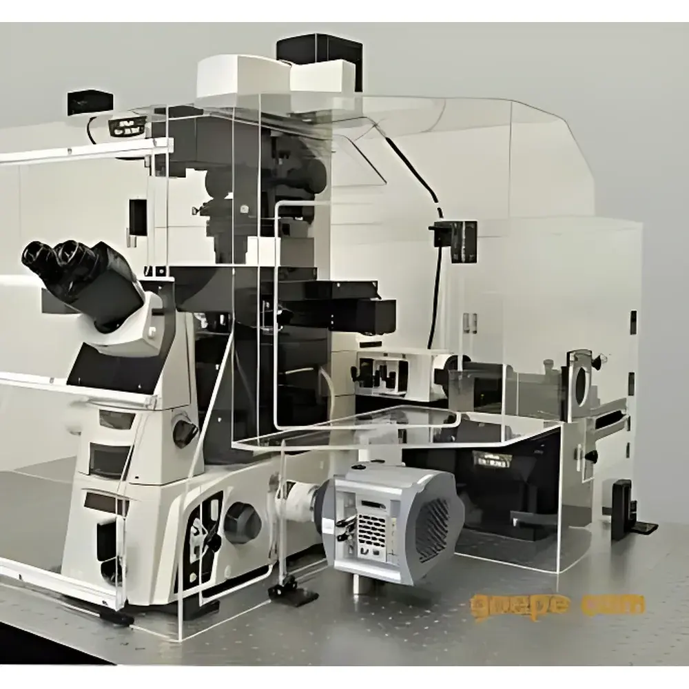
Nikon N-SIM E Super-Resolution Microscopy System
| Brand | Nikon |
|---|---|
| Origin | Japan |
| Model | N-SIM E |
| Resolution | ≤85 nm (lateral, TIRF-SIM mode) |
| Temporal Resolution | 0.6 s/frame (2D/TIRF-SIM), ~1 s/frame (3D-SIM) |
| Objective Compatibility | CFI Apo TIRF 100× Oil (NA 1.49) |
| Illumination | Multi-laser SIM (up to 5 lasers, e.g., 405/488/561/640/730 nm) |
| Imaging Modes | TIRF-SIM, 2D-SIM, 3D-SIM |
| Optical Sectioning Thickness | Up to 20 µm |
| Axial Resolution | ~300 nm (3D-SIM) |
| Compliance | Designed for GLP/GMP-aligned workflows, compatible with FDA 21 CFR Part 11–ready software platforms |
Overview
The Nikon N-SIM E Super-Resolution Microscopy System is an engineered implementation of Structured Illumination Microscopy (SIM), a widefield optical super-resolution technique that overcomes the classical Abbe diffraction limit (~200 nm lateral, ~500 nm axial) through computational reconstruction of interference patterns—Moiré fringes—generated by high-frequency sinusoidal illumination grids projected onto the specimen. Unlike single-molecule localization methods (e.g., PALM/STORM) or stimulated emission depletion (STED), SIM achieves resolution enhancement via deterministic patterned excitation and phase-shifted image acquisition, enabling quantitative, multi-color, live-cell-compatible imaging with minimal phototoxicity. The N-SIM E integrates Nikon’s proprietary optical architecture—including the CFI Apo TIRF 100× oil immersion objective (NA 1.49)—to deliver consistent lateral resolution of ≤85 nm and axial resolution of ~300 nm in 3D-SIM mode, while maintaining compatibility with standard fluorescent probes and routine cell culture protocols.
Key Features
- Diffraction-unlimited resolution: Achieves ≤85 nm lateral resolution in TIRF-SIM and 2D-SIM modes—approximately 2× improvement over conventional widefield fluorescence microscopy.
- High-speed dynamic imaging: Captures super-resolved frames at 0.6 s/frame in TIRF-SIM and 2D-SIM configurations; supports time-lapse studies of intracellular trafficking, cytoskeletal remodeling, and membrane receptor dynamics in living cells.
- Multi-modal optical sectioning: Offers three distinct operational modes—TIRF-SIM (surface-restricted, high signal-to-background), 2D-SIM (rapid whole-field super-resolution), and 3D-SIM (optical slice thickness up to 20 µm, isotropic resolution enhancement in Z).
- Multi-spectral flexibility: Compatible with the Nikon LU-5 multi-laser illumination platform, supporting up to five independently controllable solid-state lasers (e.g., 405, 488, 561, 640, and 730 nm) for simultaneous or sequential multi-channel super-resolution imaging.
- Optimized optical train: Incorporates Nikon’s CFI Apo TIRF objectives, precision motorized stage control, and thermally stabilized emission path optics to ensure repeatability across long-duration acquisitions and multi-user laboratory environments.
Sample Compatibility & Compliance
The N-SIM E system is validated for use with standard biological specimens—including adherent and suspension mammalian cells, fixed tissue sections, and live organoids—under physiological conditions (37°C, 5% CO₂). It supports common fluorophores (e.g., GFP, mCherry, Alexa Fluor dyes, SiR-tubulin) without requiring specialized photoswitching buffers. All hardware and firmware components comply with IEC 61000-6-3 (EMC emissions) and IEC 61000-6-2 (immunity) standards. When operated with Nikon’s NIS-Elements AR software configured for audit trail logging, electronic signatures, and user-access controls, the system meets foundational requirements for GLP-compliant imaging workflows and supports alignment with FDA 21 CFR Part 11 principles for regulated research environments.
Software & Data Management
Acquisition, reconstruction, and analysis are managed through Nikon’s NIS-Elements AR software platform, which provides real-time SIM reconstruction algorithms, drift correction, multi-dimensional stitching (XY, Z, time, channel), and quantitative colocalization tools (e.g., Pearson’s coefficient, Manders’ overlap). Raw image data (16-bit TIFF) and reconstructed super-resolved stacks are stored with embedded metadata—including laser power, exposure time, grating orientation, and phase shift sequences—for full experimental traceability. The software supports DICOM export for cross-platform integration and offers Python and MATLAB APIs for custom algorithm development and batch processing pipelines.
Applications
- Sub-diffraction mapping of nuclear pore complexes, centrioles, and synaptic vesicle clusters in fixed and live neurons.
- Quantitative co-distribution analysis of receptor–ligand pairs (e.g., EGFR–Grb2) at plasma membranes using dual-color TIRF-SIM.
- 3D structural dynamics of mitotic spindles and chromatin condensation during cell division.
- Long-term tracking of organelle morphology and inter-organelle contact sites (e.g., ER–mitochondria junctions) under physiological perturbations.
- Validation of CRISPR-edited protein localization phenotypes with statistical confidence exceeding conventional confocal resolution limits.
FAQ
What is the fundamental physical principle behind N-SIM’s resolution enhancement?
N-SIM exploits structured illumination—projecting known, high-spatial-frequency light patterns onto the specimen—to shift high-frequency object information into the observable passband of the optical system. Fourier-domain processing of nine raw images (three rotations × three phase shifts) enables computational recovery of fine structural detail beyond the diffraction barrier.
Can N-SIM E be used for live-cell imaging without significant photodamage?
Yes. Due to its widefield illumination geometry and relatively low peak irradiance requirements—especially in TIRF-SIM mode—the system minimizes cumulative photon dose per frame, enabling sustained imaging of sensitive primary cells and embryos over minutes to hours.
Is 3D-SIM compatible with thick tissue samples?
While optimized for monolayer and thin-sectioned specimens (≤20 µm), 3D-SIM performance degrades with increasing scattering and aberration in thicker tissues; for such applications, N-SIM E is typically paired with adaptive optics modules or cleared-tissue preparation protocols.
Does the system support third-party objective lenses?
N-SIM E is calibrated and validated exclusively for use with Nikon CFI Apo TIRF and Plan Apo λ objectives; non-Nikon lenses may compromise grating alignment fidelity and reconstruction accuracy.
How is data integrity ensured during regulatory submissions?
When deployed with NIS-Elements AR in “Compliance Mode,” the system enforces electronic signatures, immutable audit trails, role-based access control, and encrypted raw data storage—providing documentation necessary for internal QA review and external inspection readiness.


