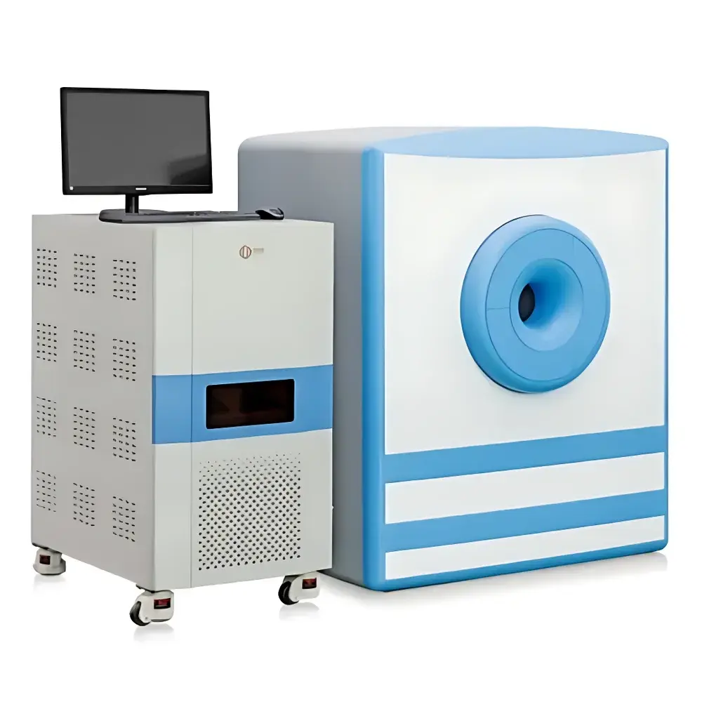
NIUMAG NM42-060H-I Animal Disease Model MRI Analyzer
| Brand | NIUMAG |
|---|---|
| Origin | Jiangsu, China |
| Manufacturer Type | Authorized Distributor |
| Country of Origin | China |
| Model | NM42-060H+ |
| Pricing | Upon Request |
| Magnetic Field Strength | 1.0 ± 0.05 T |
| Larmor Frequency | ~42 MHz |
| Animal RF Coil ID | 60 mm |
| Primary Applications | In vivo murine MRI for oncology, obesity, contrast agent development, and longitudinal disease modeling |
Overview
The NIUMAG NM42-060H-I Animal Disease Model MRI Analyzer is a compact, high-stability permanent-magnet-based benchtop magnetic resonance imaging system engineered for non-invasive, longitudinal in vivo imaging of small rodents—primarily mice and rats. Operating at a nominal static magnetic field strength of 1.0 ± 0.05 T (corresponding to a 1H Larmor frequency of approximately 42 MHz), the system leverages spin-echo and gradient-echo pulse sequences to generate quantitative anatomical and functional contrast without ionizing radiation. Unlike clinical MRI platforms, this analyzer is purpose-built for preclinical research environments where reproducibility, ease of operation, and integration into animal housing workflows are critical. Its 60 mm inner-diameter radiofrequency (RF) coil is optimized for high signal-to-noise ratio (SNR) imaging of whole-body or localized regions—including brain, subcutaneous tumors, liver, adipose depots, and orthotopic xenografts—enabling dynamic monitoring of disease progression, therapeutic response, and pharmacokinetics over time.
Key Features
- Stable permanent magnet architecture with active shimming, ensuring field homogeneity ≤ 10 ppm over a 30 mm DSV (diameter spherical volume)
- Dedicated 60 mm transmit/receive RF coil with adjustable tuning/matching for consistent excitation and reception across diverse physiological states
- Integrated gradient subsystem with maximum amplitude ≥ 200 mT/m and slew rate ≥ 1000 T/m/s, supporting diffusion-weighted imaging (DWI), perfusion mapping, and MR angiography (MRA)
- Real-time image reconstruction engine with on-board Linux-based processing unit; supports DICOM export and raw k-space data acquisition
- Modular software interface compatible with standard NIfTI and ANALYZE formats, facilitating downstream analysis in FSL, SPM, or custom MATLAB/Python pipelines
- Designed for ISO 13485-aligned manufacturing processes; CE-marked for research-use-only (RUO) applications in EU and APAC markets
Sample Compatibility & Compliance
The NM42-060H-I accommodates live mice (15–35 g) and rats (80–250 g) under isoflurane anesthesia with integrated respiratory gating and temperature regulation (37.0 ± 0.5 °C via warm-air feedback loop). Animal handling conforms to AAALAC International standards and adheres to Directive 2010/63/EU on protection of animals used for scientific purposes. All imaging protocols are documented per GLP principles, with audit trails enabled for protocol versioning, operator ID logging, and acquisition parameter stamping. While not intended for diagnostic use, the system supports method validation aligned with ASTM E2915–21 (Standard Guide for Evaluating Preclinical MRI Systems) and ISO/IEC 17025–2017 for laboratory competence in quantitative imaging biomarker development.
Software & Data Management
NIUMAG’s proprietary MREasy™ v5.x acquisition and analysis suite provides intuitive workflow-driven interface for sequence selection, parameter optimization, and real-time preview. The software enforces role-based access control (RBAC), electronic signature support, and 21 CFR Part 11-compliant audit trail generation—including timestamps, user actions, and parameter modifications—for regulated preclinical studies. Raw k-space datasets are stored in HDF5 format with embedded metadata (pulse sequence type, TR/TE, FOV, matrix size, bandwidth), enabling traceable reprocessing. Export modules support direct integration with PACS systems and cloud-based collaborative platforms such as XNAT and CBRAIN, ensuring interoperability across multi-site translational research consortia.
Applications
- Oncology: Longitudinal tracking of orthotopic glioblastoma, subcutaneous melanoma, and hepatic metastases—including tumor volume quantification, necrosis assessment via T2 mapping, and anti-angiogenic therapy response using dynamic contrast-enhanced (DCE) MRI
- Metabolic disease: Visceral vs. subcutaneous adipose tissue segmentation, hepatic steatosis grading via proton density fat fraction (PDFF), and brown adipose tissue activation mapping under cold challenge
- Neuroscience: Structural volumetry of hippocampal atrophy in Alzheimer’s transgenic models; cerebral blood volume (CBV) mapping in stroke-reperfusion paradigms
- Contrast agent development: Relaxivity (r1/r2) characterization of novel Gd-, Mn-, or iron oxide-based agents; biodistribution kinetics assessed through serial post-injection time-series
- Inflammatory disease: Detection of macrophage infiltration in colitis or arthritis models using ultrasmall superparamagnetic iron oxide (USPIO)-enhanced T2*-weighted imaging
FAQ
Is the NM42-060H-I suitable for longitudinal studies spanning multiple weeks?
Yes—the system supports daily or weekly imaging sessions with minimal thermal drift (<0.02 T/h) and automated calibration routines to maintain geometric fidelity and intensity stability across timepoints.
Can the instrument be integrated into existing vivarium infrastructure?
It features low electromagnetic emissions (EMC Class B compliance), operates on standard 230 V / 50 Hz power, and requires only 1.2 m² floor space with passive cooling—no chiller or liquid nitrogen needed.
Does NIUMAG provide application support for protocol development?
NIUMAG offers remote and on-site application scientist support, including sequence optimization workshops, phantom-based QA training, and co-development of SOPs aligned with NIH or EFPIA preclinical imaging guidelines.
What level of technical documentation is provided for regulatory submissions?
Users receive full system IQ/OQ/PQ documentation templates, traceable calibration certificates, and a comprehensive Design History File (DHF) summary upon request—supporting IND-enabling study reporting.


