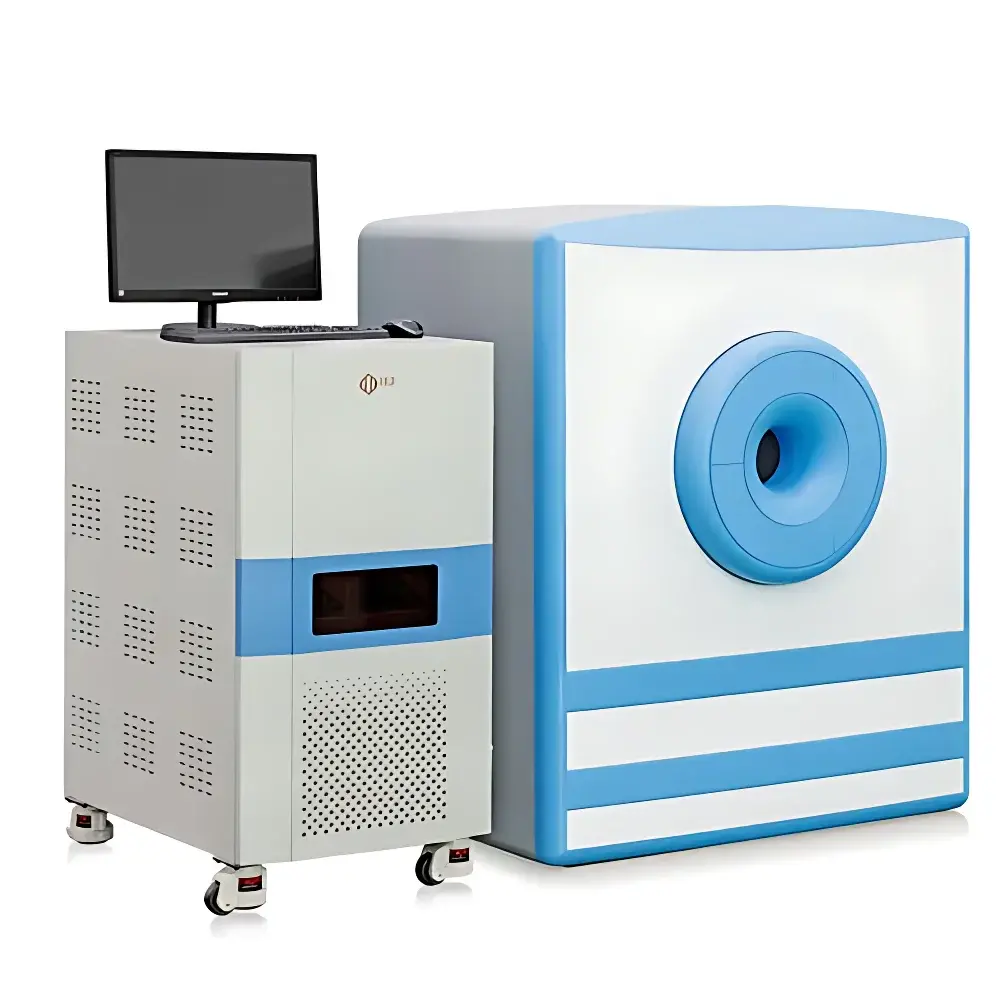
NIUMAG NM42 Small Animal MRI Scanner
| Brand | NIUMAG |
|---|---|
| Origin | Jiangsu, China |
| Manufacturer Type | Authorized Distributor |
| Country of Origin | China |
| Model | NM42-NIUMAG |
| Instrument Type | Permanent-Magnet Small Animal Magnetic Resonance Imaging System |
| Magnetic Field Strength | ~1.0 T (Permanent Magnet) |
| Bore Diameter | ≥40 mm |
| Maximum Sample Weight | 45 g |
| Minimum Sample Volume | ≥100 µL |
| Minimum Slice Thickness | 0.8 mm |
| Imaging Modes | T1-, T2-, PD-weighted, Gradient Echo, Spin Echo, Multi-slice 2D/3D acquisitions |
| Anesthesia Compatibility | Optional Integrated Gas Anesthesia System |
| Software Compliance | FDA 21 CFR Part 11–ready audit trail (optional configuration), GLP/GMP-compatible data logging |
Overview
The NIUMAG NM42 Small Animal MRI Scanner is a compact, permanent-magnet-based preclinical magnetic resonance imaging system engineered for high-fidelity, non-invasive in vivo imaging of rodents and other small laboratory animals. Operating at a stable field strength of approximately 1.0 Tesla, the NM42 leverages a high-homogeneity permanent magnet architecture to deliver robust signal-to-noise ratio (SNR), spatial resolution down to 0.8 mm slice thickness, and reproducible contrast across T1-, T2-, and proton density (PD)-weighted imaging protocols. Unlike superconducting MRI systems, the NM42 requires no liquid helium, cryogen replenishment, or RF-shielded room infrastructure—making it suitable for standard laboratory environments with standard power supply (220 V AC, 50 Hz) and ambient temperature control (18–25 °C). Its open-bore design accommodates animals up to 45 g without mechanical constraint, eliminating stress-induced physiological artifacts commonly observed in restrained or high-field systems. The scanner is fundamentally designed for longitudinal studies where repeated, low-risk imaging sessions are required—such as tumor progression monitoring, pharmacokinetic evaluation, or nanoparticle biodistribution analysis.
Key Features
- 1.0 T permanent magnet with passive shimming: Delivers field homogeneity < 10 ppm over a 30 mm DSV (diameter spherical volume), enabling high-resolution anatomical and functional imaging without cryogenic dependency.
- Non-ionizing, radiation-free imaging: Provides safe, repeatable in vivo assessment across multiple time points—critical for developmental, therapeutic, or toxicological studies compliant with IACUC and OECD guidelines.
- Three-step automated acquisition workflow: Integrated auto-tuning, center-frequency optimization, and sequence parameter selection reduce operator dependency; no advanced NMR physics expertise required.
- Modular hardware architecture: Supports optional integration of gas anesthesia delivery (isoflurane/O₂), physiological monitoring (respiratory gating, temperature feedback), and gradient coil upgrades for diffusion-weighted imaging (DWI) readiness.
- Low total cost of ownership: No cryogens, no RF shielding, minimal vibration isolation requirements, and < 1.5 kW peak power draw significantly reduce facility preparation and operational overhead.
- Open software API and DICOM 3.0 export: Enables third-party integration with PACS, ELN systems, and custom MATLAB/Python analysis pipelines.
Sample Compatibility & Compliance
The NM42 is validated for use with mice, rats, zebrafish embryos, and other small animal models with transverse dimensions ≤ 40 mm and mass ≤ 45 g. It supports both in vivo imaging and ex vivo sample analysis—including contrast agent relaxivity quantification (r₁/r₂), nanoparticle suspension characterization, microbial suspension profiling, and ion concentration mapping in aqueous media (minimum volume: 100 µL). All imaging protocols adhere to ISO/IEC 17025–aligned quality assurance practices. Optional software configurations support audit trail generation, electronic signatures, and data integrity features aligned with FDA 21 CFR Part 11 and EU Annex 11 requirements for regulated preclinical research environments.
Software & Data Management
The NM42 ships with two integrated software suites: the NIUMAG MRI Console and the NIUMAG Image Processing Suite. The Console provides full pulse sequence customization—including adjustable TR/TE, flip angle, bandwidth, and k-space sampling schemes—while maintaining intuitive graphical controls and real-time preview. Pulse sequences include spin echo, gradient echo, inversion recovery, and multi-slice turbo spin echo. The Image Processing Suite supports ROI-based quantitative analysis (T1/T2 mapping, signal intensity normalization), pseudo-color overlay, 3D volume rendering, distance/angle measurement, threshold segmentation, and batch-mode DICOM-to-NIfTI conversion. All raw k-space data and reconstructed images are stored in vendor-neutral formats (DICOM, NIfTI, MATLAB .mat) with embedded metadata (pulse sequence parameters, animal ID, date/time stamp, operator ID).
Applications
- Preclinical oncology: Longitudinal tumor volume quantification, necrosis assessment via ADC mapping, and treatment response evaluation using dynamic contrast-enhanced (DCE) MRI protocols.
- Contrast agent development: In vitro r₁/r₂ relaxivity determination per ISO 10993-18 and in vivo biodistribution tracking of Gd-based, iron oxide, or Mn-based agents.
- Neuroscience research: High-resolution brain anatomy, ventricular volume analysis, and white matter tract visualization in transgenic mouse models.
- Nanomedicine: Quantitative distribution kinetics of liposomal, polymeric, or metallic nanoparticles in liver, spleen, and tumor tissue.
- Cardiovascular phenotyping: Cine-MRI for left ventricular ejection fraction (LVEF), wall thickness, and cardiac output in murine models of hypertension or myocardial infarction.
- Musculoskeletal studies: Cartilage thickness mapping, tendon integrity assessment, and inflammatory joint volume quantification in arthritis models.
FAQ
Does the NM42 require an RF-shielded room?
No. The permanent magnet design and optimized gradient shielding eliminate the need for external RF cages or Faraday enclosures.
Can the system perform diffusion-weighted imaging (DWI)?
Yes—when equipped with the optional high-slew-rate gradient module and appropriate pulse sequence license, DWI and apparent diffusion coefficient (ADC) mapping are supported.
Is anesthesia integration mandatory?
No. While gas anesthesia (isoflurane/O₂) is recommended for motion suppression during long scans, the system supports conscious imaging for short-duration acquisitions in acclimated subjects.
What data formats are natively supported?
DICOM 3.0 (Level 3 conformance), NIfTI (.nii), and MATLAB (.mat) with full metadata embedding—including pulse sequence parameters, geometry, and acquisition timestamps.
How is system calibration maintained?
Automated daily QA routines include field homogeneity checks, RF coil tuning verification, and SNR phantoms (e.g., NiCl₂-doped water) with pass/fail reporting logged to the audit trail.


