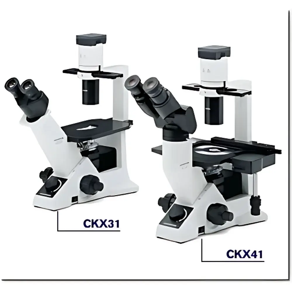
Olympus CKX31 Inverted Biological Microscope
| Brand | Olympus |
|---|---|
| Model | CKX31 |
| Optical System | UIS2 Infinity-Corrected |
| Illumination | 6V/30W Halogen Lamp with Heat-Absorbing Filter and Ground Glass Diffuser |
| Objective Turret | 4-Position Revolving Nosepiece |
| Stage | Right-Hand Low-Position Coaxial X-Y Mechanical Stage (X=120 mm, Y=78 mm) |
| Observation Tube | 30° Inclined Binocular Tube (IPD: 48–75 mm) |
| Objectives | 4× Plan Achromat, 10× Phase Contrast (PH1), 20× Long Working Distance Phase Contrast (PH1) |
| Eyepieces | 10× Widefield (FN 20) |
| Optional Accessories | Mechanical Stage Extension, Fluorescence Illuminator, CCD Imaging System |
| Compliance | Designed for ISO 9001-certified manufacturing environments |
Overview
The Olympus CKX31 Inverted Biological Microscope is engineered for routine cell culture monitoring and live-cell observation in academic, pharmaceutical, and biomanufacturing laboratories. Its inverted optical architecture positions the objective lenses beneath the specimen stage—enabling direct visualization of adherent or suspension cells within standard tissue culture vessels (e.g., flasks, Petri dishes, multi-well plates) without sample relocation or inversion. The microscope employs Olympus’ UIS2 (Universal Infinity System 2) optical platform, a fully infinity-corrected design that ensures consistent chromatic and spherical aberration correction across all magnifications. This system utilizes parallel light paths between objective and tube lens, permitting modular insertion of auxiliary components—including phase contrast sliders, fluorescence filter cubes, and digital camera adapters—without compromising image fidelity or requiring compensatory optics. With a maximum field number of 22 mm and optimized light throughput from its 6 V/30 W halogen illumination source (integrated heat-absorbing filter and ground-glass diffuser), the CKX31 delivers high-contrast, flat-field imagery from center to periphery—critical for quantitative assessment of confluency, morphology, and motility in dynamic biological systems.
Key Features
- UIS2 infinity-corrected optical pathway enabling stable resolution and minimal wavefront distortion across magnifications
- 4-position revolving nosepiece accommodating standard DIN-mount objectives (4×, 10× PH1, 20× LWD PH1)
- Ergonomic 30° inclined binocular observation tube with adjustable interpupillary distance (48–75 mm) supporting seated or standing operation
- Low-profile coaxial mechanical stage with right-hand controls and extended travel (X = 120 mm, Y = 78 mm), optimized for large-format cultureware handling
- Modular illumination base designed for rapid detachment and reconfiguration—facilitating integration into incubator-integrated imaging setups
- Phase contrast compatibility out-of-the-box, with dedicated condenser alignment and slider mechanisms for rapid PH1 optimization
- Compact footprint (W × D × H ≈ 270 × 400 × 420 mm) enabling benchtop deployment within biosafety cabinets or environmental chambers
Sample Compatibility & Compliance
The CKX31 accommodates standard upstream and downstream life science consumables: T-25/T-75 flasks, 6–96-well plates, glass-bottom dishes (e.g., MatTek, Ibidi), and chambered coverglasses. Its long-working-distance 20× phase contrast objective (≥3.5 mm clearance) permits unobstructed focusing through vessel walls up to 1.2 mm thick. All optical components meet JIS B 7151 and ISO 8578 specifications for transmitted-light microscopy performance. While the instrument itself does not carry FDA 510(k) or CE-IVD designation, it is routinely deployed in GLP-compliant cell banking, QC release testing, and preclinical assay development workflows. When paired with validated digital imaging hardware and timestamped acquisition software, the system supports traceable data capture aligned with 21 CFR Part 11 requirements for electronic records and signatures.
Software & Data Management
The CKX31 operates as a hardware platform compatible with third-party imaging suites including Olympus cellSens Dimension, MetaMorph, and open-source tools such as Fiji/ImageJ. Optional USB 3.0 CCD or CMOS cameras (e.g., DP27, SC30) provide calibrated pixel-to-micron mapping and dynamic range optimization for time-lapse quantification. Acquisition metadata—including objective ID, exposure time, gain setting, and stage coordinates—is embedded in TIFF/OME-TIFF headers. For regulated environments, audit-trail modules log user actions, parameter changes, and session timestamps—enabling retrospective verification during internal audits or regulatory inspections.
Applications
- Real-time monitoring of mammalian, insect, and stem cell cultures during expansion and differentiation protocols
- Quality control of primary cell isolates and cryopreserved vials prior to experimental use
- Assessment of transfection efficiency and reporter gene expression in adherent lines
- Pre-screening of CRISPR-edited clones based on morphological phenotypes
- Supporting automated colony picking by providing high-fidelity low-magnification overviews
- Integration into closed-loop bioreactor systems for periodic viability checks without process interruption
FAQ
Is the CKX31 suitable for fluorescence imaging?
Yes—when equipped with the optional fluorescence illuminator (e.g., U-LH100L-3) and appropriate filter sets, the CKX31 supports standard DAPI/FITC/TRITC excitation/emission bands. However, it lacks built-in shutter control or intensity modulation; external controllers are recommended for quantitative fluorescence applications.
Can the CKX31 be used inside a CO₂ incubator?
The base unit is not incubator-rated, but its modular design allows full disassembly: the stand can remain outside while only the stage and objective turret are inserted through an incubator port—provided proper thermal isolation and condensation management are implemented.
What is the maximum usable magnification with standard eyepieces?
With the supplied 10× eyepieces and 20× LWD objective, total magnification reaches 200×. Higher magnifications (e.g., 40×) require optional objectives and may reduce working distance below practical limits for vessel-based imaging.
Does the CKX31 support motorized Z-focus or stage automation?
No—focus and stage movement are manual. For automated workflows, users integrate external motorized stages (e.g., Prior ProScan II) and focus drives via TTL or RS-232 interfaces compatible with third-party acquisition software.
How does the UIS2 optical system improve image flatness compared to conventional finite systems?
By eliminating field curvature through precise correction of off-axis ray paths across the entire 22 mm field number, UIS2 achieves ≤15% improvement in edge-to-center sharpness—verified using ISO 19038 test targets under standardized Köhler illumination conditions.
Related Products