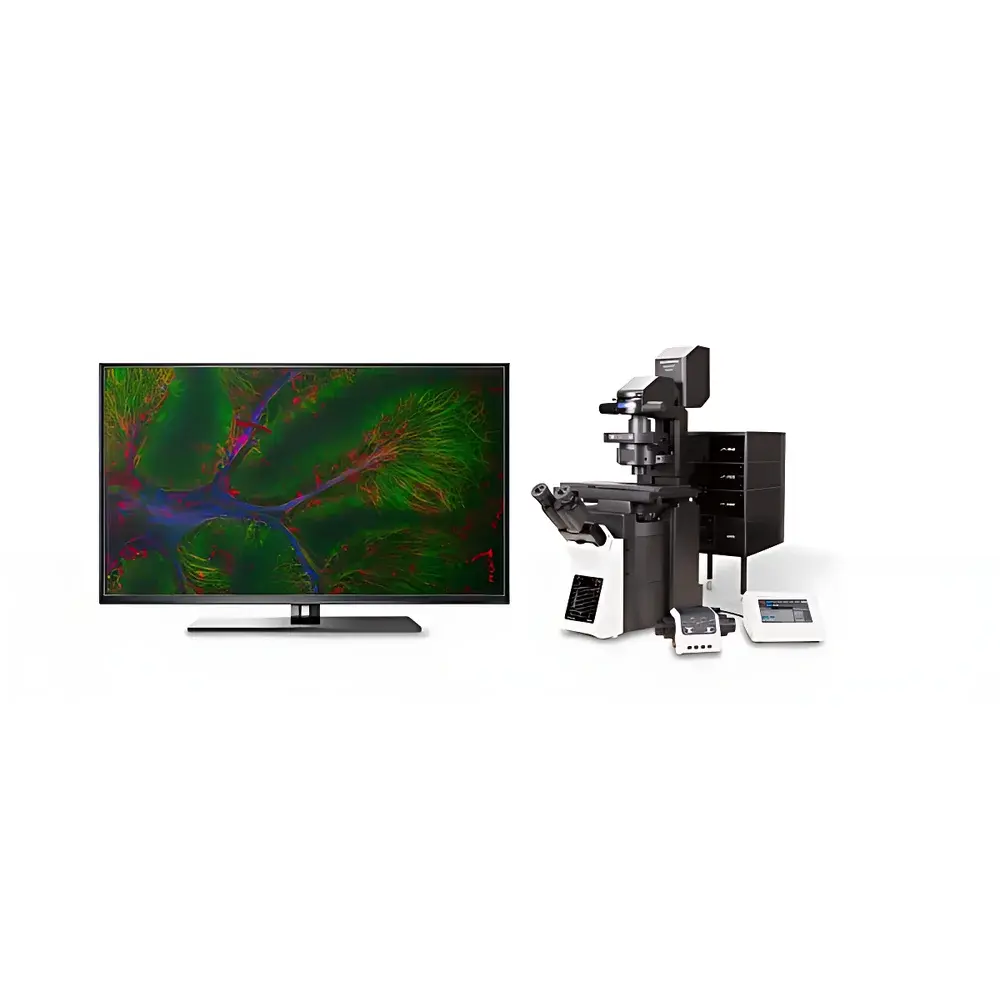
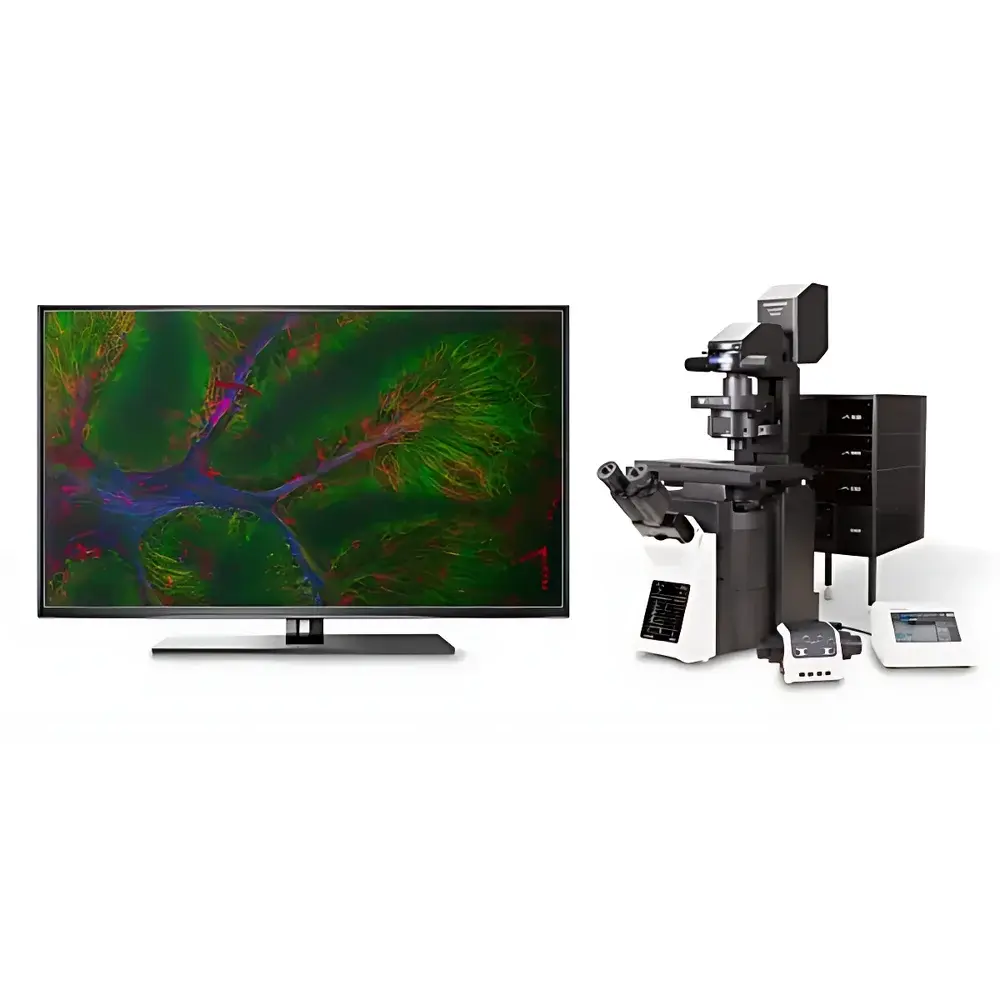
Olympus FLUOVIEW FV4000 Laser Scanning Confocal Microscope
| Brand | Olympus |
|---|---|
| Origin | Japan |
| Manufacturer Type | Original Equipment Manufacturer (OEM) |
| Product Category | Imported Instrument |
| Model | FV4000 |
| Instrument Type | Point-Scanning Confocal Microscope |
| Resolution | High Spatial and Spectral Resolution |
| Lasers | Multiple NIR-capable laser lines (405–785 nm), optional 808/980 nm for upconversion |
| Detectors | Six-channel synchronous silicon-based photodetectors (SilVIR™), 400–900 nm spectral range |
| Fluorescence Detection | SilVIR™ visible-to-infrared silicon detectors with linear response, low noise, and >6-decade dynamic range |
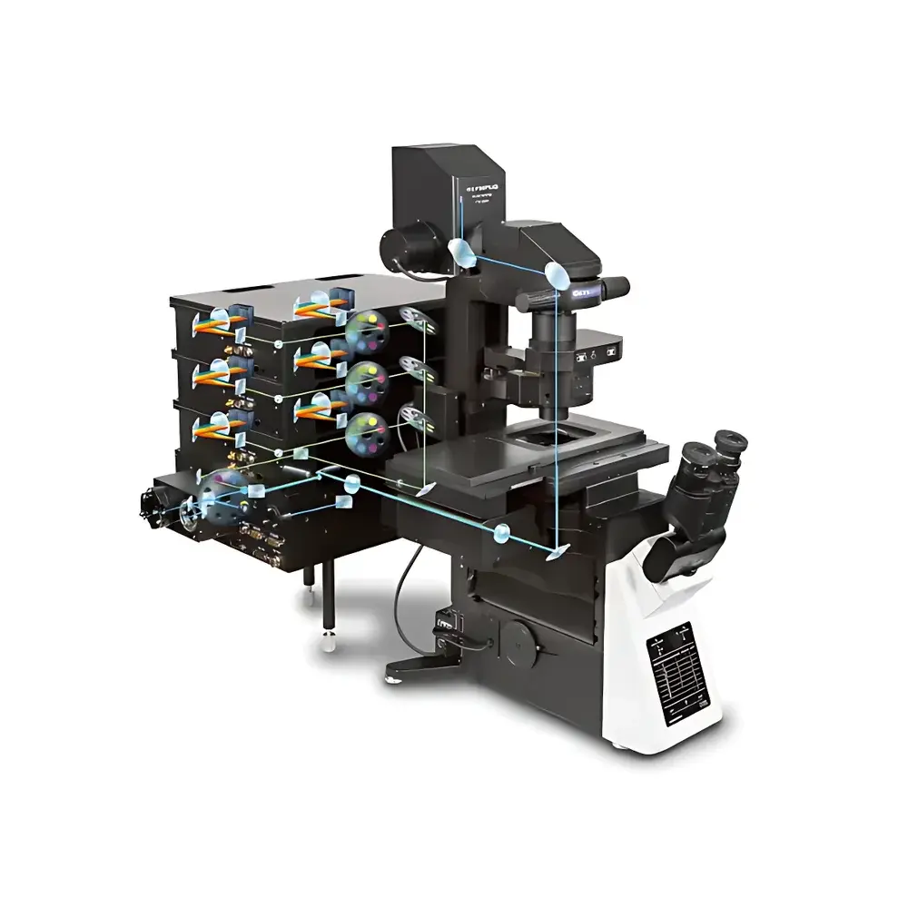
| Scanning Module | Hybrid scanning unit supporting conventional galvo + resonant scanner |
| Scan Modes | Bidirectional resonant scanning, sequential/simultaneous spectral acquisition, Z-stack, time-lapse, tile scan |
| Scan Speed | Up to 438 fps at 1024 × 32 pixels |
| Optical Zoom | Continuous digital and optical zoom with wide dynamic magnification range |
| Objectives | Wide selection of high-NA oil/water/glycerol immersion and dry objectives (e.g., UPLSAPO 60×/1.35, LUCPLFLN 60×/1.20 W) |
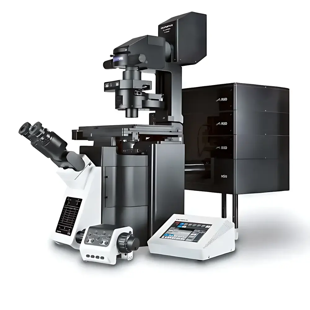
| Microscope Frame | Research-grade fully motorized inverted, upright, electrophysiology, or “gate-type” stand |
| Illumination Sources | LED-based fluorescence excitation (white LED + filter wheels), optional halogen or LED brightfield |
| Software Platform | FLUOVIEW v5.x with AI-powered denoising, automated organoid detection, C. elegans motion tracking, and multi-modal experiment scripting |
| Vibration Isolation | Dedicated active/passive anti-vibration tables (multiple footprint options) |
| XY Stage Control | Motorized stage with remote joystick and full software API integration |
Overview
The Olympus FLUOVIEW FV4000 is a point-scanning laser confocal microscope engineered for quantitative, reproducible, and spectrally resolved fluorescence imaging in life science research. It operates on the principle of spatially filtered optical sectioning—using a pinhole-confined detection path to reject out-of-focus light—and integrates advanced photonics, semiconductor detector physics, and computational imaging to achieve high-fidelity 3D reconstruction across biological scales. Unlike conventional PMT-based systems, the FV4000 leverages silicon-based SilVIR™ detectors (U.S. Patent No. US11237047), enabling linear photon counting over an extended 400–900 nm spectral window with sub-electron read noise and >6-decade dynamic range. This architecture supports rigorous quantification of fluorescence intensity, spectral unmixing, and longitudinal comparison across experiments—critical for GLP-compliant assay development, phenotypic screening, and multiplexed biomarker validation.
Key Features
- SilVIR™ Silicon Photodetectors: Six synchronized, thermoelectrically cooled silicon detectors replace traditional GaAsP-PMTs, delivering superior quantum efficiency in the near-infrared (NIR), lower dark current, and intrinsic linearity for absolute photon flux calibration.
- VPH Grating-Based Full-Spectrum Detection: Volume phase holographic (VPH) transmission gratings provide uniform spectral dispersion across all six channels, enabling 2 nm spectral resolution and 1 nm step accuracy—ideal for unmixing overlapping fluorophores without emission filter swaps.
- NIR-Optimized Optical Pathway: Custom-coated dichroics, anti-reflective optics, and corrected objective lenses maintain diffraction-limited performance from 405 nm through 785 nm, with dedicated ports for 808/980 nm upconversion excitation.
- Hybrid Resonant-Galvo Scanning: Dual-mode scanning engine supports both high-speed (438 fps at 1024 × 32) and high-resolution (1024 × 1024) acquisition, with real-time laser power feedback control ensuring consistent excitation fluence per pixel.
- AI-Enhanced Imaging Workflow: Integrated FLUOVIEW v5 software includes deep-learning-based denoising (trained on biological ground-truth datasets), automated organoid boundary segmentation, and C. elegans locomotion trajectory modeling—reducing manual ROI annotation by >70% in benchmark assays.
- Modular Expandability: Compatible with third-party environmental chambers, patch-clamp rigs, and super-resolution modules (e.g., SIM); upgrade path to FV4000MPE two-photon configuration via shared optical train and detector interface.
Sample Compatibility & Compliance
The FV4000 accommodates live-cell cultures, tissue sections (frozen or FFPE), cleared whole organs (e.g., CLARITY, iDISCO), and microfluidic devices across inverted, upright, and electrophysiology configurations. Its motorized stage and autofocus system support long-term timelapse of adherent or suspension cells under controlled CO2, temperature, and humidity. All hardware and software components comply with ISO 13485 design controls for medical device R&D environments and support FDA 21 CFR Part 11 audit trails—including electronic signatures, version-controlled acquisition protocols, and immutable raw data archiving. System validation documentation aligns with ASTM E2912 (microscope performance testing) and USP (particulate matter analysis in injectables) where applicable.
Software & Data Management
FLUOVIEW v5 provides a unified platform for experimental design, acquisition control, and quantitative analysis. The software supports OME-TIFF export compliant with Bio-Formats, HDF5-based large-volume storage for Z-stacks (>100 GB), and direct integration with Python/R via RESTful API for custom algorithm deployment. All spectral datasets are stored with embedded metadata (excitation/emission wavelengths, laser power, detector gain, objective ID) to ensure FAIR (Findable, Accessible, Interoperable, Reusable) data principles. Built-in batch processing pipelines enable automated background subtraction, spectral unmixing using reference spectra libraries, and colocalization analysis (Pearson’s r, Manders’ coefficients) with statistical confidence intervals.
Applications
- Quantitative subcellular protein localization and trafficking kinetics in live neurons and stem cell-derived organoids
- Multiplexed immunofluorescence profiling of tumor microenvironments using NIR dyes (e.g., IRDye 800CW, Alexa Fluor 750)
- Deep-tissue vascular imaging in optically cleared mouse brain using 785 nm excitation and SilVIR™ NIR detection
- Time-resolved calcium dynamics in cardiomyocyte monolayers using GCaMP6s and resonant scanning at ≥30 Hz
- Spectral fingerprinting of endogenous fluorophores (NAD(P)H, FAD, collagen SHG) in label-free tissue diagnostics
- Correlative light-electron microscopy (CLEM) workflows via precise coordinate mapping between confocal and SEM stages
FAQ
What is the spectral detection range of the SilVIR™ detectors?
The SilVIR™ detectors operate continuously from 400 nm to 900 nm, with peak quantum efficiency above 65% at 650–850 nm—significantly exceeding GaAsP-PMT performance beyond 650 nm.
Can the FV4000 be used for quantitative co-localization studies under GLP conditions?
Yes. With traceable laser power monitoring, calibrated detector linearity, and audit-trail-enabled software, the system meets requirements for quantitative colocalization in regulated preclinical studies.
Is the resonant scanner compatible with all objective magnifications?
The hybrid scanning unit maintains Nyquist-sampled resolution across 10× to 100× objectives; optimal performance is achieved with high-NA water or silicone immersion lenses optimized for NIR transmission.
Does the system support third-party software integration for machine learning analysis?
Yes. FLUOVIEW v5 exposes acquisition and metadata via documented REST API and supports direct TIFF/HDF5 export for ingestion into TensorFlow, PyTorch, or commercial platforms like Imaris or HALO.
What regulatory documentation is provided for installation qualification (IQ) and operational qualification (OQ)?
Olympus supplies IQ/OQ templates aligned with ISO/IEC 17025, including optical alignment verification, laser power stability tests, detector linearity calibration reports, and software validation summaries.


