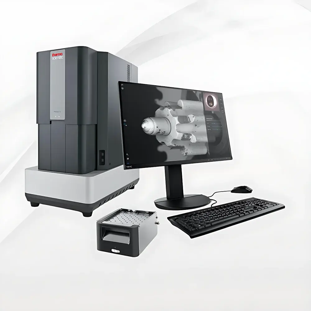
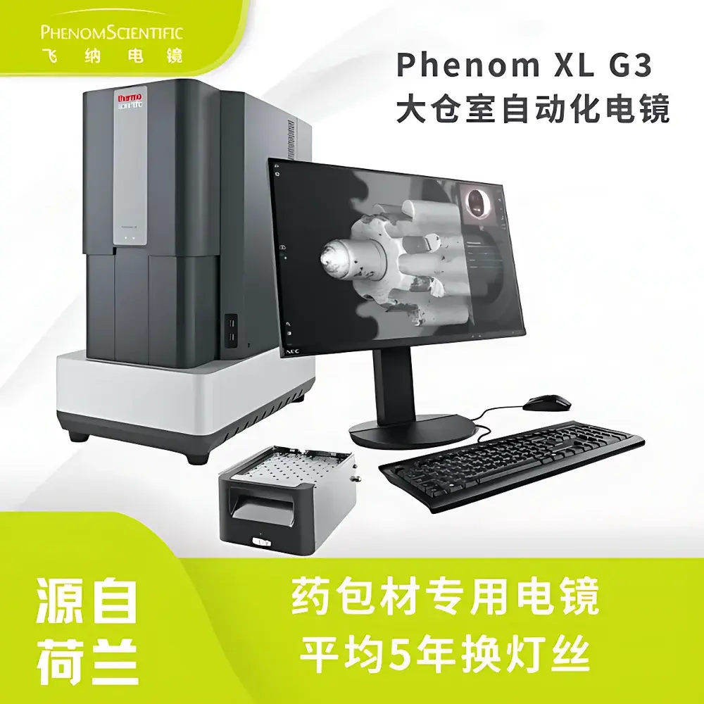
Phenom XL G3 Desktop Scanning Electron Microscope for Pharmaceutical Packaging Material Inspection
| Brand | Phenom |
|---|---|
| Origin | Netherlands |
| Model | XL G3 |
| Instrument Type | Desktop SEM |
| Electron Source | CeB6 (Cerium Hexaboride) |
| Secondary Electron Resolution | 8 nm |
| Backscattered Electron Resolution | 8 nm |
| Magnification Range | 20×–200,000× |
| Accelerating Voltage | 4.8–20.5 kV |
| Maximum Sample Diameter | 100 mm |
| Vacuum Pumping Time | <30 s |
| Optical Navigation Lens | 3–19× |
| Detector | High-Sensitivity Quad-SEG BSE Detector |
| Filament Lifetime | ~3,000 h |
| Optional | EDS Detector Integration |
Overview
The Phenom XL G3 Desktop Scanning Electron Microscope is an engineered solution for routine and regulatory-compliant microstructural analysis of pharmaceutical packaging materials, excipients, and dosage forms. Designed around a high-brightness cerium hexaboride (CeB6) thermionic electron source, the XL G3 delivers stable beam current and superior signal-to-noise ratio—enabling consistent 8 nm secondary and backscattered electron (SE/BSE) resolution across its full magnification range (20× to 200,000×). Unlike conventional tungsten-filament SEMs, the CeB6 source provides higher brightness at lower operating voltages, reducing charging artifacts on insulating samples such as polymer films, blister packs, and lyophilized matrices—without mandatory metal coating. Its compact desktop architecture integrates a large 100 mm sample chamber, permitting direct observation of intact vials, ampoules, pouches, and tablet blister strips in their native state. The system achieves vacuum readiness in under 30 seconds via integrated turbomolecular pumping, eliminating the need for dedicated vibration-isolated rooms or HVAC modifications. This design aligns with the technical requirements outlined in the draft 2025 edition of the Chinese Pharmacopoeia, where SEM-based particulate characterization—including insoluble particle morphology, foreign contaminant identification, and surface defect mapping—is formally codified for packaging integrity assessment.
Key Features
- High-stability CeB6 electron source with ~3,000-hour operational lifetime and low energy spread—optimized for high-resolution imaging of non-conductive pharmaceutical substrates
- Integrated optical navigation lens (3–19×) enabling rapid area selection and seamless transition from macro to micro-scale observation
- Quad-segmented backscattered electron (BSE) detector supporting atomic number contrast imaging, phase differentiation, and semi-quantitative compositional mapping
- Full automation of vacuum cycling, focus, stigmation, and brightness/contrast—requiring no user intervention for routine inspection workflows
- Vibration-resistant mechanical design certified for operation in standard laboratory, QC, or manufacturing environments (no active damping required)
- Modular upgrade path to energy-dispersive X-ray spectroscopy (EDS), supporting elemental identification per USP <1087>, ISO 13485, and ICH Q5E guidelines
Sample Compatibility & Compliance
The XL G3 accommodates diverse pharmaceutical specimens without destructive preparation: uncoated polymer films (e.g., PVC/PVDC/aluminum laminates), freeze-dried cakes, gelatin capsules, suspension droplets dried on conductive stubs, and filter-captured insoluble particles (≥1 µm). Its low-voltage imaging capability (down to 4.8 kV) minimizes beam-induced damage to heat-sensitive or volatile components. All imaging protocols—including stage positioning, dwell time, and detector gain settings—are fully documented and exportable to support GLP/GMP audit trails. System software complies with FDA 21 CFR Part 11 requirements for electronic records and signatures when configured with role-based access control and audit logging. Method validation documentation—including resolution verification per ASTM E1558, charge mitigation performance on insulators, and repeatability testing per ISO/IEC 17025—is available upon request for regulatory submissions.
Software & Data Management
Phenom’s proprietary ProSuite software provides intuitive, workflow-driven operation with preconfigured application templates for pharmaceutical use cases: “Particle Morphology,” “Blister Seal Integrity,” “Coating Uniformity,” and “Foreign Material ID.” Image metadata—including acquisition parameters, calibration timestamps, operator ID, and instrument serial number—is embedded in TIFF and JPEG exports. Raw image data is stored in vendor-neutral formats compatible with third-party image analysis platforms (e.g., ImageJ/Fiji, MATLAB, or commercial particle analyzers). For enterprise integration, the system supports DICOM export, network file sharing (SMB/NFS), and API-based automation via Python SDK for batch processing and LIMS synchronization.
Applications
- Visual identification and morphological classification of insoluble particles in injectables and ophthalmic solutions per USP <788> and <789>
- Surface topography analysis of primary packaging materials (e.g., crimp seal defects, laminate delamination, pinhole detection)
- Morphometric evaluation of granules, pellets, and tablets—including porosity, coating thickness uniformity, and erosion patterns
- Contaminant forensics: differentiation of silicone oil droplets, stainless steel wear debris, glass fragments, or cellulose fibers based on BSE contrast and optional EDS signature
- Stability study support: longitudinal monitoring of packaging degradation (e.g., oxidation-induced surface cracking, moisture ingress pathways)
FAQ
Does the XL G3 meet the requirements specified in the draft 2025 Chinese Pharmacopoeia for SEM-based packaging inspection?
Yes—the system satisfies all referenced technical criteria, including CeB6 source specification, resolution verification protocol, and sample handling methodology for uncoated insulators. Documentation packages for pharmacopoeial alignment are provided with installation.
Can I analyze hydrated or volatile samples without critical-point drying?
The XL G3 is not a variable-pressure or environmental SEM; therefore, fully hydrated samples require prior fixation and drying. However, it routinely images dried residues from aqueous suspensions, lyophilized powders, and solvent-evaporated films without charging artifacts.
Is EDS integration validated for elemental quantification in regulated environments?
EDS add-on modules include factory-calibrated standards and SOP templates aligned with ISO 14707 and ASTM E1508. Quantitative accuracy is verified using NIST-traceable reference materials, and full validation reports are supplied for IQ/OQ/PQ execution.


