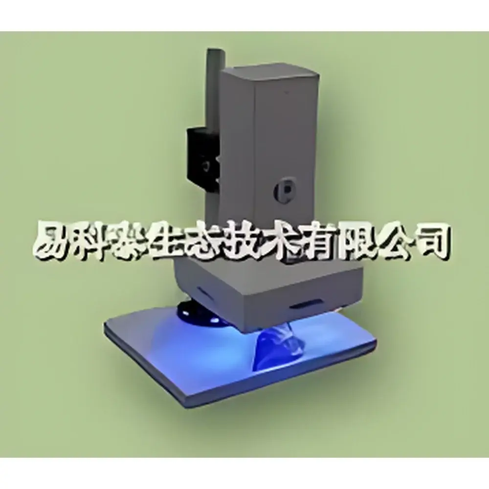
Photon Systems Instruments FC 1000-H/GF Portable GFP & Chlorophyll Fluorescence Imaging System
| Origin | Europe |
|---|---|
| Manufacturer Type | Authorized Distributor |
| Import Status | Imported |
| Model | FC 1000-H/GF |
| Pricing | Available Upon Request |
| Illumination Area | 3.5 × 3.5 cm |
| Measuring Light Wavelengths | 620 nm (red), 455 nm (blue) |
| Measuring Light Pulse Duration | 10–250 µs |
| Actinic Light (Blue) | ~350 µmol·m⁻²·s⁻¹ |
| Saturating Pulse (Blue) | up to 1,000 µmol·m⁻²·s⁻¹ |
| Far-Red Light | 735 nm |
| CCD Sensor | 512 × 512 pixels (standard), optional 640 × 480 or 1392 × 1040 |
| Pixel Size | 8.2 × 8.4 µm |
| Spectral Range | 400–1000 nm |
| Quantum Efficiency | 70% at 540 nm, 50% at 400 nm and 650 nm |
| Read Noise | <12 e⁻ RMS (typ. 10 e⁻) |
| Full Well Capacity | >70,000 e⁻ (unbinned) |
| Frame Rate | 50 fps |
| A/D Resolution | 12-bit |
| Power Supply | 90–260 V AC or sealed lead-acid battery |
| System Weight (main unit) | 1.8 kg |
| Tripod Weight | 1.5 kg |
| Power Module Weight | 2.5 kg |
| Total Portable Setup Weight (incl. laptop & accessories) | ~3.5 kg |
| Max Power Consumption | 200 W |
| Communication Interface | USB 2.0 |
| Firmware | Upgradable via BIOS |
Overview
The Photon Systems Instruments FC 1000-H/GF is a field-deployable, dual-modality fluorescence imaging system engineered for quantitative spatiotemporal analysis of chlorophyll a fluorescence and green fluorescent protein (GFP) expression in intact plant tissues and small organisms. Based on the well-established FC 1000-H platform, the H/GF variant integrates optimized optical pathways and synchronized LED excitation sources to enable simultaneous or sequential acquisition of both physiological (photosynthetic) and molecular (transgene expression) fluorescence signals. Its measurement principle relies on high-speed, time-resolved detection of transient fluorescence kinetics—including the Kautsky induction curve, non-photochemical quenching (NPQ), and photochemical quenching (qP)—using pulsed measuring light and precisely timed actinic and saturating pulses. The system captures two-dimensional fluorescence parameter maps across a defined 3.5 × 3.5 cm field of view, delivering pixel-level quantification of over 50 biophysically relevant parameters such as F₀, Fₘ, Fᵥ/Fₘ, ΦPSII, NPQ, ETR, qN, qP, and Rfd. Designed for minimal perturbation and maximal reproducibility, it supports both laboratory-controlled experiments and uncontrolled field conditions—making it suitable for longitudinal studies under natural irradiance, drought, temperature stress, or pathogen challenge.
Key Features
- Dual-channel capability: Simultaneous or alternating imaging of chlorophyll fluorescence (ChlF) and GFP emission without optical filter swaps
- Modular LED illumination system: Four high-intensity, uniform LED panels (4 × 4 cm) with independently controllable red (620 nm), blue (455 nm), and far-red (735 nm) sources
- Programmable light regimes: User-defined actinic light intensity (0–350 µmol·m⁻²·s⁻¹), saturating pulse amplitude (up to 1,000 µmol·m⁻²·s⁻¹), and temporal sequence via embedded scripting language
- High-sensitivity scientific CCD: 512 × 512 pixel sensor (8.2 × 8.4 µm pixels), 12-bit digitization, quantum efficiency >70% at 540 nm, read noise <12 e⁻ RMS
- Field-ready architecture: Integrated sealed lead-acid battery operation, lightweight carbon-fiber tripod (1.5 kg), shoulder-carry case, and ruggedized housing rated for outdoor use
- Firmware-upgradable BIOS and USB 2.0 interface ensure long-term compatibility and remote diagnostics support
Sample Compatibility & Compliance
The FC 1000-H/GF accommodates a broad range of biological specimens including detached leaves, whole seedlings (<15 cm height), algal biofilms, cyanobacterial colonies, fruit surfaces, and small model organisms (e.g., Arabidopsis thaliana, Brachypodium distachyon, Chlamydomonas reinhardtii). Its compact imaging area and adjustable working distance allow precise targeting of heterogeneous tissue zones—such as stomatal clusters, lesion margins, or root-shoot junctions—without sample dissection. All fluorescence parameters are calculated in accordance with internationally accepted conventions (Strasser et al., 2004; Maxwell & Johnson, 2000) and align with ASTM E2919-13 (Standard Guide for Chlorophyll Fluorescence Measurements in Plants) and ISO 10211:2019 (Plant physiology — Measurement of photosynthetic performance). Data acquisition workflows support GLP-compliant metadata tagging, timestamped audit trails, and export formats compatible with FDA 21 CFR Part 11–enabled LIMS environments when integrated with validated FluorCam software configurations.
Software & Data Management
FluorCam v7.x (Windows 10/11 compatible) provides an intuitive graphical interface with preloaded standard protocols—including rapid light curves (RLCs), OJIP transients, and dark-adapted Fᵥ/Fₘ screening—as well as a built-in macro language (FluorScript) for custom experimental logic. The software enables automatic ROI detection and segmentation across multiple samples within a single frame, batch processing with barcode scanner integration, and kinetic modeling of fluorescence decay profiles. Raw image stacks (16-bit TIFF) and derived parameter maps are stored with embedded EXIF-style metadata (exposure time, LED intensities, ambient temperature, GPS coordinates if enabled). Export options include Excel (.xlsx), CSV, HDF5, and MATLAB-compatible .mat files. All processing steps—including background subtraction, flat-field correction, and parameter normalization—are fully traceable and reproducible, satisfying requirements for method validation under ISO/IEC 17025 and GMP Annex 11.
Applications
- Transgenic line screening: Spatial mapping of GFP-tagged protein localization and expression dynamics under developmental or stress-induced promoters
- Photosynthetic phenotyping: High-throughput assessment of PSII quantum yield, electron transport rate (ETR), and photoprotective capacity across mutant libraries or breeding populations
- Biotic and abiotic stress diagnostics: Early detection of drought, salinity, heavy metal toxicity, or pathogen infection via altered NPQ kinetics and Fᵥ/Fₘ depression gradients
- Stomatal heterogeneity analysis: Correlation of spatially resolved ΦPSII patterns with gas exchange measurements and epidermal anatomy
- Plant-microbe interactions: Quantification of symbiotic or pathogenic colonization effects on host photosynthetic efficiency and redox status
- Fruit ripening and postharvest physiology: Monitoring chlorophyll degradation kinetics and associated shifts in fluorescence lifetime signatures
FAQ
Can the FC 1000-H/GF operate without AC power in remote field locations?
Yes—the system is fully battery-operated using its integrated sealed lead-acid power module, enabling continuous acquisition for up to 4 hours per charge under typical measurement protocols.
Is calibration required before each experiment?
No routine recalibration is needed; however, daily flat-field correction using the included reference panel is recommended for optimal radiometric consistency across environmental conditions.
Does the software support automated multi-timepoint experiments?
Yes—FluorCam allows scheduling of unattended time-series acquisitions with configurable intervals, environmental logging, and conditional triggers based on fluorescence thresholds.
Are spectral filters included for GFP-specific detection?
The system includes factory-aligned bandpass filters optimized for GFP emission (500–550 nm) and chlorophyll fluorescence (680–740 nm); additional filter sets are available as optional accessories.
How is data integrity ensured during long-term deployments?
All raw images and processed outputs include cryptographic hash verification, version-stamped firmware logs, and optional encrypted storage modes compliant with NIST SP 800-171 data protection standards.


