PSI FluorCam Large-Scale Plant Chlorophyll Fluorescence and Multispectral Imaging Platform
| Brand | PSI (Czech Republic) |
|---|---|
| Origin | Czech Republic |
| Model | FluorCam Large-Scale Plant Chlorophyll Fluorescence and Multispectral Imaging Platform |
| Imaging Area | 35 × 35 cm |
| CCD Resolution | 1360 × 1024 pixels |
| Frame Rate | up to 20 fps at full resolution |
| A/D Depth | 16-bit (65,536 gray levels) |
| Pixel Size | 6.45 µm × 6.45 µm |
| Excitation Wavelengths | 320–400 nm (UV-A), 450 nm (blue), 620 nm (red), white light, 735 nm (NIR) |
| Filter Wheel | 7-position motorized |
| Adjustable Imaging Height | 350–1350 mm |
| Compliance | ASTM E2912, ISO 17025-compatible workflow support, GLP/GMP-ready data audit trail (via software timestamping and protocol versioning) |
Overview
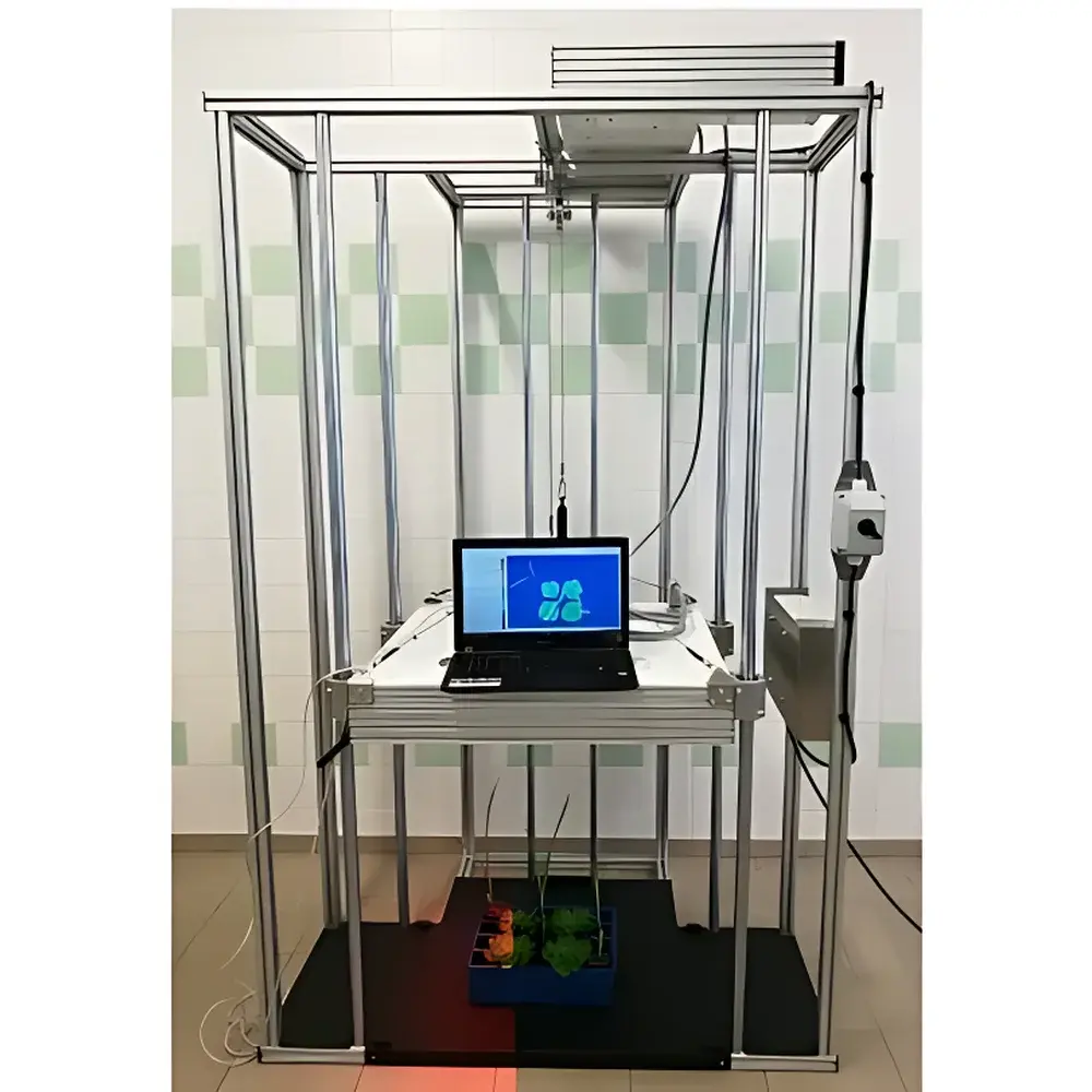
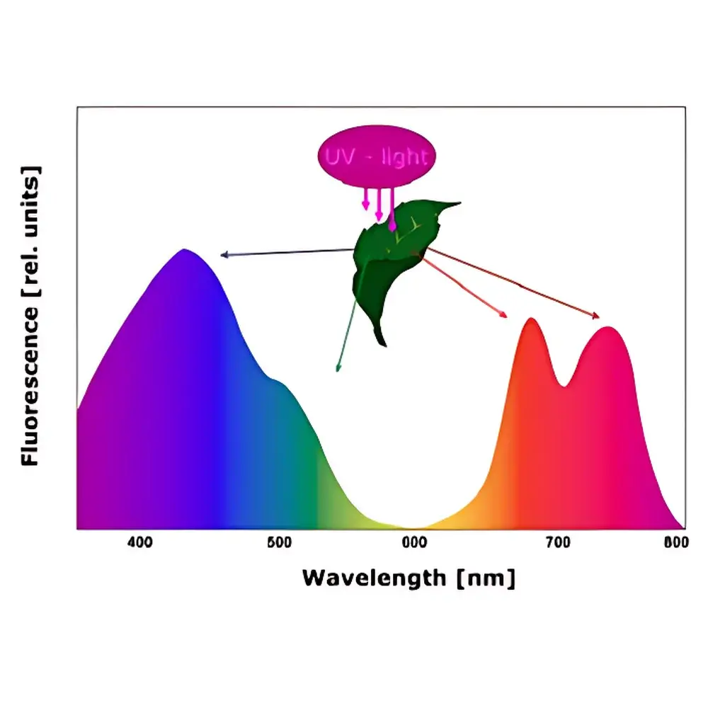
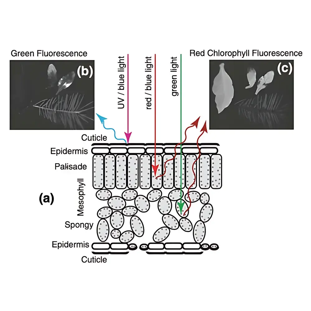
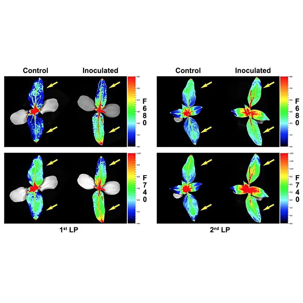
The PSI FluorCam Large-Scale Plant Chlorophyll Fluorescence and Multispectral Imaging Platform is a high-precision, non-invasive phenotyping instrument engineered for quantitative functional imaging of plant physiological status across spatial and temporal scales. Based on the physical principles of chlorophyll fluorescence induction kinetics and multispectral excitation-emission spectroscopy, the system captures dynamic photochemical responses in vivo by measuring photon emission following controlled optical excitation. It operates under two primary modalities: (1) modulated chlorophyll fluorescence imaging using red (620 nm) and far-red (735 nm) actinic and saturating pulses—enabling calculation of PSII quantum yield (ΦPSII), non-photochemical quenching (NPQ), and photochemical quenching (qP); and (2) UV-A (320–400 nm) and visible-light-excited multispectral fluorescence imaging, resolving four diagnostic emission bands: F440 (blue), F520 (green), F690 (red), and F740 (far-red). These bands originate from distinct biochemical compartments—cell wall lignins and flavonoids (F440/F520), and photosystem II antenna complexes (F690/F740)—providing orthogonal metrics for stress detection, metabolic profiling, and phenotypic trait quantification.
Key Features
- World’s largest single-frame fluorescence imaging area (35 × 35 cm), enabling whole-plant or multi-plant cohort analysis without image stitching or geometric distortion.
- Motorized 7-position filter wheel with user-selectable excitation/emission bandpass filters, supporting dual- or triple-channel sequential imaging (e.g., GFP + Chl-F + UV-induced BGF).
- Programmable LED illumination array (750 × 750 mm) with independent control of red (620 nm), blue (450 nm), white, NIR (735 nm), and UV-A (365 nm) sources—each calibrated for irradiance uniformity (±5% across field-of-view).
- High-sensitivity cooled CCD camera with 16-bit digitization, 20 fps video mode for kinetic analysis, and snapshot mode optimized for stable fluorophore detection (e.g., GFP, YFP, DAPI).
- Automated height-adjustable imaging gantry (350–1350 mm working distance) with precision linear actuator and position feedback—ensuring consistent magnification and focus across heterogeneous plant architectures.
- Integrated PAR absorption and NDVI imaging module (680 nm + 735 nm LEDs + matched filters) for concurrent structural-functional assessment.
- Optional IR thermography unit (640 × 512 uncooled microbolometer, 7.5–13.5 µm spectral range, ±0.03 °C thermal sensitivity) co-registered with fluorescence data for integrated water-use efficiency and stomatal conductance inference.
Sample Compatibility & Compliance
The platform accommodates diverse biological specimens including intact seedlings, mature rosettes, herbaceous and woody stems, detached leaves, flowers, fruits, roots, algae suspensions, and small model organisms (e.g., Arabidopsis, maize, tomato, barley, Chlamydomonas). Sample mounting is flexible—no vacuum or adhesive required—and compatible with standard growth trays, Petri dishes, multi-well plates, and custom-stage fixtures. Data acquisition workflows adhere to Good Laboratory Practice (GLP) requirements: all measurements are time-stamped, protocol versions are logged, raw kinetic traces are retained in lossless TIFF format, and software supports 21 CFR Part 11-compliant electronic signatures when deployed in regulated environments. Instrument performance is traceable to NIST-calibrated photodiode standards; factory calibration certificates accompany each delivery.
Software & Data Management
FluorCam Control & Analysis Software (v8.x) provides an integrated environment for experimental design, real-time monitoring, preprocessing, statistical mapping, and report generation. Core modules include: Live Mode (for hardware diagnostics and manual parameter tuning), Protocol Editor (with drag-and-drop scripting interface supporting conditional logic, nested loops, and variable-duration light steps), Pre-processing Engine (supporting automated ROI detection via template matching, manual polygonal selection, multi-threshold segmentation, and morphological masking), and Results Dashboard (generating parametric heatmaps, kinetic curves per ROI, histogram distributions, and export-ready Excel/CSV tables). Advanced features include “signal-then-average” vs. “average-then-signal” processing modes for optimal SNR management, batch processing of unlimited samples with auto-ID recognition, and synchronized multi-modal data fusion (fluorescence + thermal + RGB). All metadata—including light intensities, exposure times, filter positions, and environmental logs—are embedded in image headers and preserved through export.
Applications
This platform serves as a core infrastructure tool in academic, breeding, and regulatory laboratories for: drought and salinity stress phenotyping; early detection of biotic stressors (pathogens, herbivory); nitrogen use efficiency screening; heavy metal phytotoxicity assessment; UV-B acclimation studies; genetic mapping of photosynthetic traits; high-throughput mutant screening; seed vigor and germination kinetics; ecological toxicology (e.g., pesticide mode-of-action studies); and validation of remote sensing algorithms. Its capacity to resolve both photochemical (Fv/Fm, ΦPSII) and structural-metabolic (F440/F520 ratio, NDVI, thermal heterogeneity) parameters within a single measurement cycle enables systems-level interpretation of plant performance under complex environmental gradients.
FAQ
What is the maximum plant height supported for full-field imaging?
The gantry accommodates plants up to 100 cm tall while maintaining optimal working distance (350–1350 mm) and uniform illumination across the 35 × 35 cm FOV.
Can the system quantify absolute fluorescence intensity, or only relative ratios?
While absolute radiometric calibration is available as an optional add-on (using NIST-traceable reference standards), standard operation reports normalized, unitless fluorescence parameters (e.g., Fv/Fm, NPQ) derived from standardized induction protocols—ensuring inter-laboratory reproducibility.
Is remote operation and long-term unattended monitoring supported?
Yes. The system supports scheduled autonomous runs (up to two interleaved protocols), local storage (SSD-based), network streaming via GigE Vision, and integration with LIMS or farm-management platforms via RESTful API.
How does the software handle heterogeneous sample batches during automated analysis?
ROI detection uses adaptive thresholding combined with shape-prior templates (e.g., circular for petri dishes, rectangular for trays); users may define custom masks or apply machine-learning-assisted segmentation in post-processing.
Are firmware and software updates provided after purchase?
PSI offers free minor-version software updates for the duration of hardware warranty (2 years); major-version upgrades and extended support contracts are available under annual maintenance agreements.
Related Products


