RELION CL-MicroSpectra Cathodoluminescence-Enhanced Microspectroscopy System
| Brand | RELION |
|---|---|
| Origin | USA |
| Manufacturer Type | Authorized Distributor |
| Import Status | Imported |
| Model | RELION CL-MicroSpectra |
| Operating Principle | Electron Beam–Induced Cathodoluminescence Spectroscopy |
| Imaging Modality | Computerized Tomographic Spectral Mapping |
| Platform Compatibility | Ground-Based & Airborne Integration |
| Spectral Range | 200–850 nm (CL Emission), Extended to 1–10,000 µm² spatial sampling area |
| Spectral Resolution | 1–15 nm (FWHM) |
| Spatial Resolution (IFOV) | 1 nm (at optimal beam focus) |
| Field of View (TFOV) | 8 mm diameter |
| Imaging Frame Rate | 15 fps (full-frame spectral cube acquisition) |
| Vacuum Chamber | ≤0.25 Pa base pressure |
| Electron Gun | Cold Cathode, Horizontal Geometry |
| Acceleration Voltage | 0–30 kV (adjustable in 100 V increments |
| Beam Current | 0.15–1 mA continuous (5 mA peak, current-limited with thermal feedback) |
| Spot Size Control | Variable demagnification from defocused (≥5 µm) to diffraction-limited point focus (<10 nm at 25 kV) |
| Digital Interface | Real-time vacuum, HV, beam current, and operational mode display with status logging |
Overview
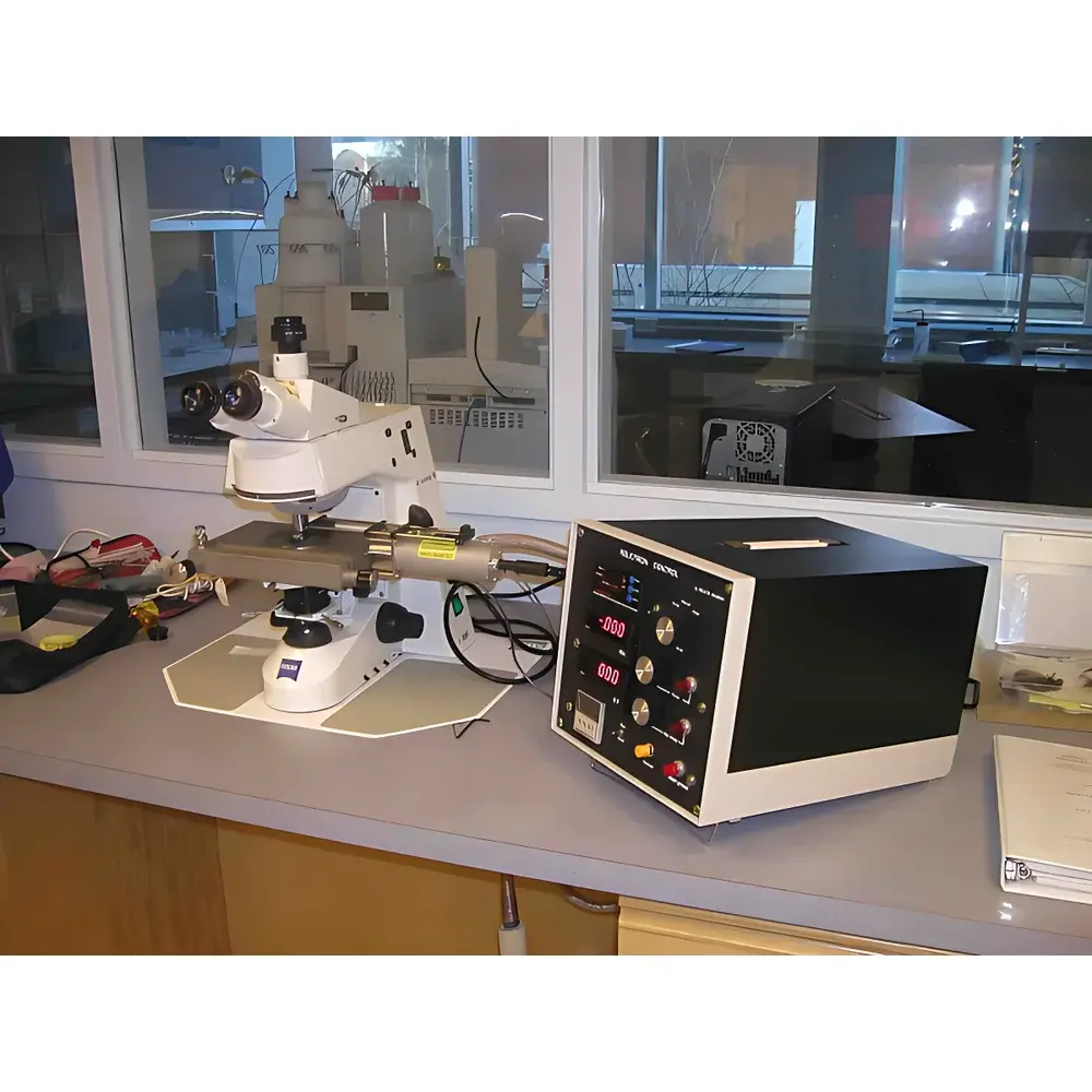
The RELION CL-MicroSpectra Cathodoluminescence-Enhanced Microspectroscopy System is a high-precision analytical platform engineered for correlative microstructural and optical spectroscopic characterization of inorganic solids at sub-micron spatial resolution. It integrates a high-stability cold-cathode electron gun with a vacuum-compatible optical coupling interface into standard optical or scanning electron microscopes (SEM-CL), enabling cathodoluminescence (CL) excitation across the ultraviolet–visible–near-infrared range (200–850 nm). Unlike conventional photoluminescence or laser-excited techniques, CL leverages high-energy electron bombardment (5–25 kV) to generate deep-volume excitation (up to ~10 µm penetration depth in dielectrics such as quartz or calcite), producing spectrally rich emission signatures tied directly to crystal lattice defects, trace-element doping, strain fields, and bandgap transitions. The system captures full spectral cubes—spatially registered CL intensity maps across wavelength—with 1 nm instantaneous field-of-view (IFOV), 1–15 nm spectral resolution, and real-time 15 fps frame acquisition. Its design adheres to ASTM E1557 (Standard Guide for Cathodoluminescence Analysis of Geological Materials) and supports GLP-compliant data provenance through timestamped metadata embedding.
Key Features
- Vacuum-integrated cold-cathode electron column: Operates at ≤0.25 Pa base pressure, preserving sample integrity during extended CL acquisition; compatible with cryogenic stages for low-temperature CL studies.
- Variable-beam excitation optics: Electron acceleration voltage (0–30 kV) and current (0.15–1 mA) are digitally stabilized and logged; spot size continuously adjustable from broad-area irradiation to nanoscale probing via electromagnetic focusing.
- Modular spectral detection architecture: Supports interchangeable grating modules (150–1200 grooves/mm) and back-illuminated CCD/CMOS detectors, enabling optimization for UV sensitivity, NIR extension (to 1100 nm), or Raman-ready configurations (with 532/785/1064 nm laser ports).
- Co-registered imaging-spectroscopy workflow: Combines wide-field optical microscopy (TFOV = 8 mm) with diffraction-limited spectral sampling (IFOV = 1 nm); CMS optical path switcher enables seamless toggling between broadband imaging, monochromatic CL mapping, and multi-band spectral acquisition.
- Hardware-synchronized data acquisition: All spectral frames are time-stamped and tagged with concurrent vacuum pressure, beam parameters, and stage coordinates—enabling retrospective audit trails compliant with FDA 21 CFR Part 11 requirements.
Sample Compatibility & Compliance
The RELION CL-MicroSpectra is validated for analysis of polished thin sections, bulk mineral fragments, semiconductor wafers, geological drill cores, and ceramic composites. Its non-destructive excitation mechanism preserves sample morphology and chemical integrity—critical for archival specimens and regulatory submissions. Sample chamber accommodates standard 25 × 75 mm microscope slides and custom mounts up to 50 mm diameter. The system meets ISO/IEC 17025:2017 clause 5.9 (measurement traceability) when paired with NIST-traceable spectral calibration standards (e.g., Hg-Ar lamp, Si reference). For pharmaceutical or forensic applications, it supports USP analytical instrument qualification (AIQ) protocols, including operational qualification (OQ) templates for beam stability, spectral repeatability (<±0.3 nm drift over 4 h), and spatial registration accuracy (<±50 nm over 1 mm scan).
Software & Data Management
Acquisition and processing are managed via RELION SpectraSuite v4.x—a modular, Python-extendable platform supporting batch spectral deconvolution, multivariate curve resolution (MCR), and principal component analysis (PCA). Raw hyperspectral data are stored in HDF5 format with embedded EXIF-like metadata (beam energy, dwell time, grating position, detector gain). SpectraSuite includes built-in compliance tools: electronic signature capture, 21 CFR Part 11–compliant audit trail (user actions, parameter changes, file exports), and role-based access control (RBAC) for multi-user labs. Export options include CSV, JCAMP-DX, and Bruker OPUS-compatible formats for cross-platform spectral library integration (e.g., RRUFF, Mindat.org). Optional add-ons enable automated mineral phase classification using convolutional neural networks trained on >12,000 labeled CL spectra from sedimentary, igneous, and metamorphic systems.
Applications
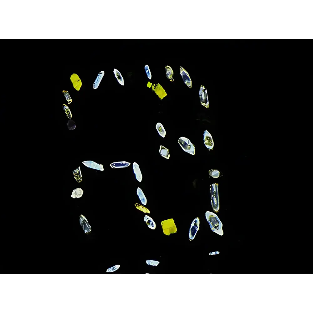
- Geological Petrochronology: Discrimination of detrital quartz provenance via CL zonation patterns; quantification of calcite cement growth sequences in sandstone pore networks; identification of authigenic vs. diagenetic feldspar based on Mn²⁺/Fe³⁺-activated emission bands.
- Materials Science: Mapping dislocation density in GaN epitaxial layers via CL peak broadening; correlating grain boundary segregation in perovskite solar cell absorbers with localized non-radiative recombination centers.
- Forensic & Cultural Heritage: Non-invasive pigment stratigraphy in historical paintings; differentiation of synthetic vs. natural gemstones (e.g., sapphire vs. ruby) using Cr³⁺/Fe²⁺-sensitive CL line ratios.
- Microelectronics: Defect localization in SiC power devices; assessment of radiation damage in nuclear-grade ceramics via CL lifetime decay kinetics (time-resolved CL module optional).
FAQ
What vacuum level is required for stable CL operation?
The system achieves optimal signal-to-noise ratio at ≤0.25 Pa base pressure, maintained by a dual-stage turbomolecular pump; no liquid nitrogen cooling is required for standard operation.
Can the RELION CL-MicroSpectra be retrofitted to existing optical microscopes?
Yes—vacuum interface adapters are available for Zeiss Axio, Leica DM series, and Nikon Eclipse platforms; mechanical clearance ≥45 mm between objective and stage is required.
Is spectral calibration traceable to national standards?
All factory calibrations use NIST SRM 2035 (Hg-Ar emission lamp) and SRM 2036 (Si reflectance standard); calibration certificates include uncertainty budgets per GUM guidelines.
Does the system support time-resolved CL measurements?
Time-resolved capability (100 ps–10 µs decay lifetimes) is available via optional pulsed electron gun upgrade and TCSPC detector module.
How is data integrity ensured during long-duration acquisitions?
Real-time checksum validation, cyclic redundancy checks (CRC-32), and automatic write-verification ensure bit-perfect HDF5 file generation; all metadata are cryptographically signed upon export.
Related Products


")