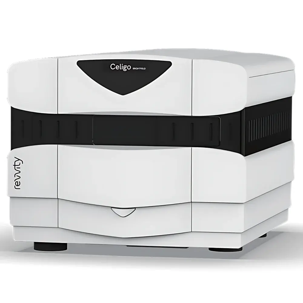
Revvity Celigo Whole-Well Cell Imaging Cytometer
| Brand | Revvity |
|---|---|
| Origin | USA |
| Manufacturer Type | Original Equipment Manufacturer (OEM) |
| Product Origin | Imported |
| Model | Celigo |
| Pricing | Available Upon Request |
Overview
The Revvity Celigo Whole-Well Cell Imaging Cytometer is a high-throughput, automated digital imaging platform engineered for label-free and fluorescence-based quantitative analysis of adherent and suspension cells directly within standard microplates and culture vessels. Operating on wide-field brightfield and multi-channel fluorescence microscopy principles, the Celigo captures full-well, tile-scanned images without requiring cell detachment or staining—enabling non-invasive, longitudinal monitoring of cellular phenotypes across time. Its optical architecture integrates precision motorized stage control, high-resolution CMOS imaging sensors, and calibrated LED excitation sources to ensure consistent illumination and spatial fidelity across 6- to 1536-well plates and T-flasks. Designed for integration into regulated life science workflows, the system delivers reproducible, objective metrics—including confluence, cell count, size distribution, morphology parameters, and fluorescence intensity per object—without reliance on subjective manual scoring.
Key Features
- Whole-well imaging with auto-focus and adaptive exposure optimization per well and channel
- Simultaneous acquisition across one brightfield channel and up to four independent fluorescence channels (e.g., DAPI, FITC, TRITC, Cy5)
- High-speed scanning: complete imaging of a 96-well plate in under 8 minutes; 384-well in under 15 minutes
- Automated multi-timepoint acquisition with configurable intervals for kinetic growth, cytotoxicity, or migration assays
- Integrated gating and population segmentation algorithms enabling flow-like analysis of heterogeneous cell populations based on morphology and intensity thresholds
- Real-time image overlay capability: merge brightfield with fluorescent channels to visualize subpopulation localization within monolayers or 3D structures
- Rugged mechanical design with temperature- and CO2-compatible enclosure options for on-stage incubation
Sample Compatibility & Compliance
The Celigo supports diverse biological sample formats including adherent monocultures, co-cultures, suspension cells, spheroids, organoids, iPSC-derived colonies, and tumor stem cell clusters. It accommodates standard ANSI/SLAS-compliant microplates (6–1536-well), Petri dishes, and T-25/T-75 flasks without adapter modification. All image acquisition and analysis protocols are fully traceable and support audit-ready documentation. The system complies with GLP and GMP-aligned data integrity requirements, including user access controls, electronic signatures, and full audit trail logging per FDA 21 CFR Part 11 guidelines. Image metadata conforms to MIAME and MIAPE reporting standards where applicable.
Software & Data Management
Celigo’s proprietary software operates on a workflow-driven interface with drag-and-drop protocol building, eliminating scripting requirements for routine assays. Each analysis module—including Confluence Analysis, Cell Counting, Cytotoxicity (e.g., LDH, Caspase-3), Apoptosis (Annexin V/PI), Migration (scratch/wound healing), Invasion (Matrigel-coated transwell), and Phosphorylation (p-ERK/p-AKT quantification)—is pre-validated and parameter-tunable. Raw TIFF and JPEG2000 image files are stored with embedded EXIF metadata. Quantitative outputs export to CSV, Excel, or structured XML for LIMS integration. Optional API access enables bidirectional communication with ELN systems and statistical platforms such as GraphPad Prism or R.
Applications
The Celigo platform is routinely deployed in academic research, biopharmaceutical development, and contract research organizations for applications including: primary cell expansion monitoring; CRISPR clone isolation and validation; CAR-T manufacturing QC; 3D tumor spheroid drug response profiling; stem cell differentiation kinetics; receptor internalization kinetics; phagocytosis efficiency assays; and high-content screening of kinase inhibitors. Its ability to quantify rare events—such as low-frequency iPSC colonies or apoptotic bodies in heterogeneous cultures—makes it particularly valuable in regenerative medicine and oncology target discovery pipelines.
FAQ
Does the Celigo require cell labeling for basic confluence or counting assays?
No—brightfield imaging enables accurate, label-free confluence measurement and morphology-based cell identification across most adherent lines.
Can Celigo analyze cells in thick 3D matrices like collagen gels or Matrigel?
Yes, using extended depth-of-field (EDF) scanning and z-stack reconstruction modules, the system resolves objects up to ~200 µm in thickness with maintained segmentation accuracy.
Is instrument calibration traceable to NIST standards?
Yes—system-level photometric and spatial calibration is performed using NIST-traceable reference slides and documented in the Certificate of Conformance supplied with each unit.
How does Celigo handle focus drift during long-term time-lapse experiments?
It employs real-time autofocus correction at every timepoint using contrast-based algorithms and optional hardware-based focus lock sensors.
Are analysis protocols compliant with regulatory submissions (e.g., IND, BLA)?
Yes—software validation packages, IQ/OQ/PQ documentation, and 21 CFR Part 11 compliance modules are available upon request for regulated environments.
Related Products


