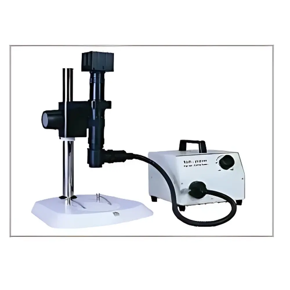
SPM Single-Tube Polarizing Microscope
| Origin | Beijing |
|---|---|
| Manufacturer Type | Distributor |
| Origin Category | Domestic |
| Model | SPM |
| Price | USD 1,390 (FOB) |
| Magnification Range | 0.7×–4.5× continuous zoom (6.42:1 zoom ratio) |
| Objective Series Optical Magnification | 0.21×–360× |
| Working Distance | 30 mm–331 mm |
| Standard Configuration | 1× objective lens, 1× CCD eyepiece |
| Tube Diameter | Ø39 mm |
| Main Unit Length | 200 mm |
| Working Distance with 1× Objective | 96 mm |
| Planar Measurement Stage | 25 mm × 25 mm, resolution 0.01 mm |
| Eyepiece-Camera Adapter Interface | C-mount |
| Illumination | Coaxial fiber-optic light source with integrated polarizer/analyzer control |
| Adjustable Aperture Diaphragm | Steel-blade type, positioned between eyepiece and zoom body |
| Crosshair Reticle | Integrated between eyepiece and C-mount, displayed superimposed on live imaging output |
| Manual 3D Rotational Mount | 360° continuous rotation at objective end |
Overview
The SPM Single-Tube Polarizing Microscope is an engineered solution for routine and research-grade birefringence analysis, crystallographic orientation assessment, and anisotropic material characterization in industrial quality control and academic laboratories. Built upon a monolithic single-tube optical architecture, it eliminates alignment drift associated with compound-tube systems while preserving high axial stability and polarization fidelity. Its core optical design follows the principles of transmitted and reflected polarized light microscopy—employing strain-free optics, precision-aligned linear polarizers, and rotatable analyzer modules—to enable quantitative evaluation of retardation, extinction angles, and interference color patterns per ASTM E112 and ISO 10810 standards. The system operates in both brightfield and polarized contrast modes, supporting identification of crystalline phases, polymer spherulites, mineral inclusions, and stress-induced birefringence in transparent or semi-transparent specimens.
Key Features
- Single-tube coaxial optical path: Minimizes mechanical misalignment, ensures consistent polarization plane integrity across magnification changes, and enhances long-term repeatability in comparative studies.
- Continuously variable zoom optics (0.7×–4.5×, 6.42:1 ratio): Coupled with interchangeable objective lenses (0.21×–360× total magnification range), enabling seamless transition from macro-scale sample survey to microstructural detail without refocusing or tube reconfiguration.
- Coaxial fiber-optic illumination module: Integrates a high-color-rendering LED light engine with independently adjustable polarizer and analyzer elements—reducing stray light, improving contrast-to-noise ratio, and facilitating extinction angle calibration.
- Steel-blade aperture diaphragm: Positioned between the eyepiece and zoom body to precisely regulate effective numerical aperture—increasing depth of field without compromising resolution, especially critical for thick or layered anisotropic samples.
- C-mount compatible crosshair reticle: Optically embedded between the eyepiece and camera interface; delivers stable, parfocal alignment of measurement fiducials with real-time imaging output—essential for coordinate-based metrology and documentation workflows.
- Manual 360° rotational mount at objective nose: Enables true three-dimensional angular inspection of specimens—supporting oblique-view birefringence mapping, cleavage plane identification, and multi-angle extinction analysis beyond conventional vertical-axis constraints.
Sample Compatibility & Compliance
The SPM accommodates standard microscope slides (26 mm × 76 mm), petrographic thin sections (30 µm thickness), bulk polymer films, ceramic fracture surfaces, and metallurgical mounts up to 331 mm working distance. Its optical train complies with ISO 8578 (microscope mechanical tube length tolerances) and incorporates strain-free lens elements certified per DIN EN 61000-6-3 for electromagnetic compatibility in shared lab environments. Polarization components meet ISO 10110-5 specifications for wavefront distortion (< λ/4 @ 550 nm). While not intrinsically GLP/GMP validated, the system supports audit-ready documentation when paired with compliant imaging software—enabling traceable image capture, timestamped metadata logging, and user-access-controlled calibration records required under FDA 21 CFR Part 11 Annex 11 frameworks.
Software & Data Management
The SPM interfaces seamlessly with third-party machine vision platforms (e.g., HALCON, OpenCV-based custom applications) and commercial microscopy suites (e.g., NIS-Elements, Stream Motion). Its C-mount output delivers native 1920×1080 video stream with 8-bit grayscale or RGB Bayer encoding. Optional SDKs provide low-level access to exposure time, gain, white balance, and trigger synchronization—facilitating automated polarizer rotation sequences and time-lapse retardation mapping. All captured images retain EXIF-compliant metadata including objective magnification, polarizer angle, aperture setting, and system timestamp—ensuring full experimental provenance for peer-reviewed publication or internal QA reporting.
Applications
- Geological thin-section analysis (feldspar twinning, quartz c-axis orientation, mica pleochroism)
- Polymer processing QC (crystallinity mapping, phase separation in blends, residual stress in injection-molded parts)
- Pharmaceutical solid-state characterization (polymorph differentiation, hydrate/anhydrate identification, tablet coating uniformity)
- Materials science research (liquid crystal domain structure, ferroelectric domain wall dynamics, composite fiber alignment)
- Forensic fiber analysis (synthetic fiber birefringence profiling, natural vs. artificial fiber discrimination)
- Electronics failure analysis (stress-induced birefringence in encapsulants, solder joint anisotropy)
FAQ
Does the SPM support motorized polarizer rotation?
No—the current configuration uses manual polarizer and analyzer controls. Motorized rotation is available as a factory-installed option (SPM-MPR) with programmable angular resolution of 0.1° and RS-232/USB command interface.
Is the 0.01 mm stage resolution verified per ISO 230-2?
Yes—the XY translation stage undergoes factory calibration using laser interferometry; certificate of conformity (CoC) is supplied with each unit.
Can the system be adapted for fluorescence observation?
Not natively—the optical path lacks dichroic beam splitters or excitation filter slots. However, external epi-illumination modules with polarization-preserving optics may be integrated via side-port adapters.
What is the maximum specimen height clearance with the 1× objective?
At 96 mm working distance, the system accommodates specimens up to 85 mm in height when using the standard base plate; extended-height stages are available as accessories.
Is DIC (Differential Interference Contrast) compatible?
No—DIC requires specialized Wollaston prisms and Nomarski condensers not supported by this optical architecture. Polarization contrast remains the primary contrast mechanism.


