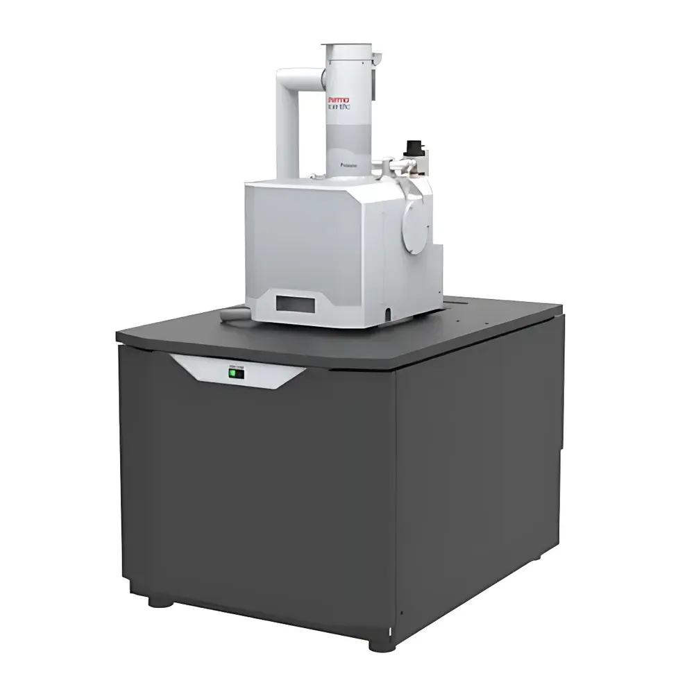
Thermo Fisher Prisma E SEM – Environmental Scanning Electron Microscope with Tungsten Filament
| Brand | Thermo Fisher |
|---|---|
| Origin | Czech Republic |
| Model | Prisma E SEM |
| Instrument Type | Floor-standing SEM |
| Electron Source | Tungsten Filament |
| Secondary Electron Resolution | 3.0 nm @ 30 kV (SE), 8.0 nm @ 3 kV (SE) |
| Backscattered Electron Resolution | 4.0 nm @ 3 kV (BSE), 10 nm @ 3 kV (SE) |
| Accelerating Voltage | 200 V – 30 kV |
| Magnification Range | 5× – 1,000,000× |
| Low Vacuum Range | up to 130 Pa |
| ESEM Mode Pressure | up to 2600 Pa (H₂O), 4000 Pa (N₂) |
| Sample Chamber Width | 340 mm |
| Expandable Ports | 12 total, including 3 EDS ports (2 at 180° opposing positions) |
| Stage | 5-axis motorized eucentric stage (X=110 mm, Y=110 mm, Z=65 mm, T=–15° to 90°, R=360° continuous) |
| Standard Detectors | ETD (high-vacuum SE), LVD (low-vacuum SE), GSED (ESEM SE), IR CCD camera in chamber |
Overview
The Thermo Fisher Prisma E SEM is a floor-standing environmental scanning electron microscope engineered for high reproducibility and operational flexibility across diverse vacuum regimes — from high vacuum (HV) to low vacuum (LV) and full environmental scanning electron microscopy (ESEM) conditions. Its core architecture integrates a thermionic tungsten filament electron source with a robust, dual-anode electron optical column optimized for stability and beam current control (up to 2 µA, continuously adjustable). Unlike field-emission instruments, the Prisma E SEM delivers consistent performance for routine and multi-user laboratory environments where reliability, ease of maintenance, and broad sample compatibility are prioritized over ultra-high resolution at cryogenic or ultra-high vacuum extremes. The system operates across an accelerating voltage range of 200 V to 30 kV, enabling both surface-sensitive low-kV imaging and deeper material interaction at higher energies. Its ESEM capability — supported by water vapor or nitrogen partial pressures up to 2600 Pa (H₂O) or 4000 Pa (N₂) — permits direct observation of hydrated, insulating, or outgassing specimens without conductive coating, preserving native morphology and chemical state.
Key Features
- Multi-regime vacuum operation: seamless switching between high vacuum (≤10⁻³ Pa), low vacuum (up to 130 Pa), and ESEM mode (up to 2600 Pa H₂O), eliminating the need for sputter coating of non-conductive or volatile samples.
- 5-axis fully motorized eucentric stage with precise navigation: X/Y/Z linear travel (110/110/65 mm), tilt (–15° to +90°), and continuous 360° rotation — enabling tilt-series acquisition, cross-sectional analysis, and automated multi-point workflows.
- Integrated Nav-Cam™ optical navigation system: a built-in IR-CCD camera inside the specimen chamber provides real-time macroscopic context and Montage-based coordinate mapping, significantly reducing time-to-target for heterogeneous or large-area samples.
- Dedicated detector suite: high-efficiency Everhart-Thornley detector (ETD) for HV SE imaging; low-vacuum detector (LVD) for charge-free LV imaging; gas secondary electron detector (GSED) optimized for ESEM signal amplification under controlled gas environments; plus in-chamber IR-CCD for positional verification and thermal monitoring.
- ColorSEM™ technology integration: enables simultaneous acquisition and real-time overlay of elemental distribution maps (via optional EDS) onto topographic SEM images — supporting rapid qualitative phase identification without interrupting imaging sessions.
- Expandable platform architecture: 12 standardized flange ports on the specimen chamber, including three dedicated EDS ports (two positioned at diametrically opposed 180° locations for optimal solid angle and shadowing minimization), facilitating straightforward integration of STEM-in-SEM, cathodoluminescence, heating/cooling stages, or micro-manipulators.
Sample Compatibility & Compliance
The Prisma E SEM is designed for laboratories requiring regulatory-compliant, repeatable imaging of industrially and biologically relevant specimens. Its ESEM functionality supports ASTM E2727-18 (Standard Guide for ESEM Use in Materials Characterization) and aligns with ISO/IEC 17025 requirements for method validation in testing laboratories. The system accommodates samples up to 340 mm wide, 200 mm tall, and 5 kg in mass — compatible with standard 12 mm pin mounts (up to 18 simultaneously) or custom fixtures. Uncoated biological tissues, polymers, ceramics, geological sections, and battery electrode cross-sections can be imaged in near-native states. For GLP/GMP environments, the Windows 10-based control software supports user access levels, audit trail logging, and electronic signature compliance per FDA 21 CFR Part 11 when configured with appropriate IT infrastructure.
Software & Data Management
Control and acquisition are managed via Thermo Fisher’s proprietary SmartSEM™ software, running on a dedicated workstation equipped with a 24-inch 1920×1200 LCD display. The GUI supports four-panel real-time image viewing, customizable toolbars, and context-sensitive workflow wizards. All imaging parameters — including dwell time, scan speed, contrast/brightness, stigmation, and detector gain — are stored with metadata in TIFF or BMP format. The software includes full Undo/Redo functionality for parameter adjustments and annotation tools compliant with ISO 14971 risk management documentation. Raw data export supports HDF5 and EMF formats for third-party processing (e.g., DigitalMicrograph, ImageJ/Fiji, or MATLAB). Automated report generation templates adhere to ISO/IEC 17025 documentation standards, including instrument ID, operator credentials, calibration timestamps, and environmental chamber logs.
Applications
- Materials science: failure analysis of uncoated composites, porosity quantification in catalysts, grain boundary characterization in sintered oxides, and in situ thermal expansion studies using integrated heating stages.
- Life sciences: imaging of freeze-dried or critical-point-dried plant tissues, fungal hyphae, insect cuticles, and soft hydrogels — all without metal coating or dehydration artifacts.
- Geosciences: examination of clay mineral aggregates, pore-network mapping in shale cores, and fluid inclusion morphology under controlled humidity.
- Electronics & energy: inspection of Li-ion battery electrodes pre- and post-cycling, solder joint integrity, and semiconductor packaging delamination — leveraging low-kV imaging to minimize charging and beam damage.
- Forensics & quality control: fiber identification, paint layer stratigraphy, and wear debris analysis — enabled by rapid sample loading, intuitive navigation, and traceable imaging protocols.
FAQ
What vacuum modes does the Prisma E SEM support, and how do they differ?
The system operates in three distinct vacuum regimes: high vacuum (≤10⁻³ Pa) for maximum resolution and EDS sensitivity; low vacuum (up to 130 Pa) for moderate charge dissipation on insulators; and ESEM mode (up to 2600 Pa H₂O) for true environmental imaging of wet or outgassing samples using gas ionization signal amplification.
Is the tungsten filament compatible with long-term, high-throughput lab use?
Yes — the dual-anode thermionic source offers predictable lifetime (>100 h at typical operating currents), simplified replacement procedures, and stable emission characteristics ideal for shared-core facilities where uptime and technician accessibility are critical.
Can the Prisma E SEM be upgraded with EDS or other analytical modules post-purchase?
Absolutely — the 12-port chamber design and standardized flange interfaces (CF35/CF63) allow field-installation of EDS detectors, WDS spectrometers, or STEM detectors without chamber venting or major reconfiguration.
Does the system meet regulatory requirements for pharmaceutical or medical device QA/QC?
When deployed with validated software configurations, electronic audit trails, and documented calibration records, the Prisma E SEM supports compliance with ISO 13485, USP , and FDA 21 CFR Part 11 for analytical instrument qualification in regulated environments.
What is the practical utility of the 340 mm chamber width?
This dimension enables direct mounting of large-format substrates (e.g., 8-inch wafers, composite panels, or geological thin sections), eliminates frequent sample repositioning, and supports multi-region correlation workflows — such as linking optical microscopy fields-of-view to targeted SEM sites via Nav-Cam™ coordinates.


