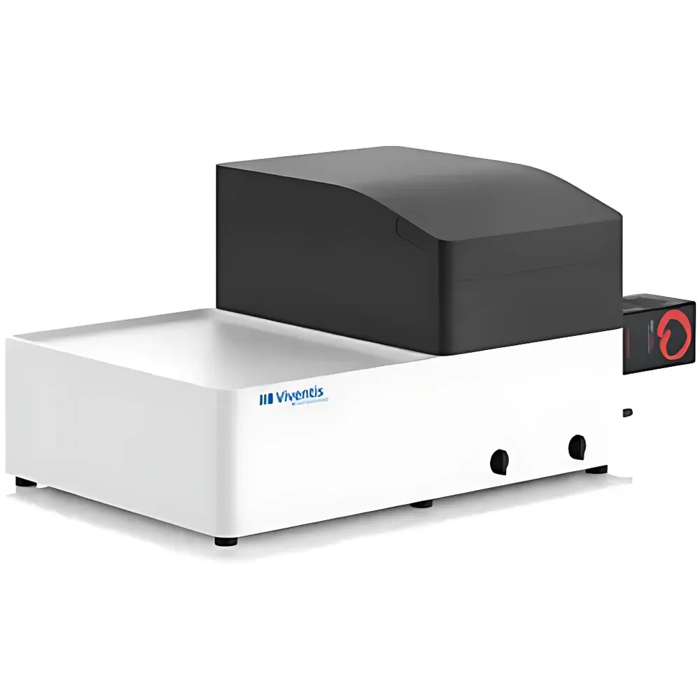
Viventis LS2 Long-Term High-Resolution Organoid Light Sheet Microscope
| Brand | Viventis Microscopy |
|---|---|
| Origin | Switzerland |
| Model | LS2 |
| Light Sheet Technology | Dual-side illumination with scanned Gaussian light sheet |
| Imaging Resolution | 406 nm (16× objective), 260 nm (25× objective) |
| Field of View | 900 µm (16×), 596 µm (25×) |
| Maximum Sample Size | >50 mm |
| Stage Travel Range | 50 mm |
| Positioning Repeatability (±) | <100 nm in XYZ |
| Laser Wavelengths | 405 nm, 488 nm, 561 nm, 638 nm |
| Objectives | Dual 25× water-immersion, NA 1.1 |
| Camera | Dual high-sensitivity sCMOS sensors, QE ≥80%, acquisition speed >30 fps |
| Illumination | Dual-side illumination |
Overview
The Viventis LS2 Long-Term High-Resolution Organoid Light Sheet Microscope is a purpose-built platform engineered for quantitative, low-phototoxicity volumetric imaging of delicate, light-sensitive living specimens—including mammalian oocytes, early embryos, cerebral and intestinal organoids, tumor microtissues, and zebrafish embryos. Unlike conventional widefield or confocal microscopy, the LS2 employs orthogonal light sheet illumination: a thin, scanned Gaussian light sheet (1.5–6 µm thickness, software-tunable and auto-calibrated) intersects the detection axis perpendicularly, illuminating only the focal plane. This geometry minimizes out-of-plane excitation, drastically reducing photobleaching and photodamage while enabling sustained time-lapse acquisition over hours to days. The system integrates dual high-NA water-immersion objectives (25×, NA 1.1) and dual sCMOS cameras (QE ≥80%, >30 fps), delivering sub-260 nm lateral resolution at 25× magnification—validated against ISO 19012-1 standards for optical sectioning performance. Its open architecture supports precise synchronization of illumination, stage motion, and camera triggering, making it suitable for GLP-compliant longitudinal studies in developmental biology and regenerative medicine.
Key Features
- Dual-side scanned Gaussian light sheet generation with real-time software control of beam translation, rotation, and thickness—enabling adaptive optical sectioning across heterogeneous sample geometries.
- Ultra-stable XYZ positioning stage with <100 nm repeatability and 50 mm travel range, optimized for long-term drift compensation during multi-hour acquisitions.
- Modular laser engine supporting four fixed wavelengths (405/488/561/638 nm), Class 3B certified, with optional custom wavelength integration for spectral unmixing or optogenetic compatibility.
- Large-format sample chamber accommodating specimens up to >50 mm in diameter—compatible with embedded organoids in Matrigel® or synthetic hydrogels, as well as multi-well embryo arrays.
- Dual sCMOS detection path with independent gain, exposure, and ROI control per camera—enabling simultaneous multi-angle acquisition or rapid Z-stack reconstruction without mechanical repositioning.
- Integrated autofocus and region-of-interest (ROI) targeting via detection-path laser reflection—facilitating rapid navigation and reproducible sub-volume imaging in opaque or scattering samples.
Sample Compatibility & Compliance
The LS2 is validated for imaging intact, unperturbed biological systems ranging from single-cell embryos to 3D organoid cultures (>300 µm thick) and tumor co-cultures (e.g., fibroblast–cancer cell spheroids). Its low-dose illumination protocol adheres to the ALARA principle (As Low As Reasonably Achievable) for live-sample integrity, satisfying requirements for IACUC- and ethics-board-approved longitudinal experiments. Hardware and firmware comply with CE marking directives (2014/30/EU EMC, 2014/35/EU LVD) and ISO 13485:2016 quality management frameworks. Optional audit-trail logging and user-access controls support alignment with FDA 21 CFR Part 11 for regulated preclinical imaging workflows. All optical components are traceably calibrated using NIST-traceable microsphere standards, and resolution verification follows ASTM E2867–21 guidelines for light sheet microscope characterization.
Software & Data Management
The LS2 is operated via Viventis AcquireSuite™—a modular, Python-extendable platform designed for reproducible experiment definition and execution. It features intuitive workflow templates for common assays (e.g., organoid growth kinetics, embryonic axis formation, mitotic spindle dynamics), with one-click switching between single-plane, multi-position, or multi-timepoint modes. Raw data is saved in standardized OME-TIFF format with embedded metadata (acquisition parameters, stage coordinates, laser power, objective ID), ensuring FAIR (Findable, Accessible, Interoperable, Reusable) compliance. Built-in GPU-accelerated deconvolution (Richardson–Lucy algorithm) and real-time background subtraction reduce post-processing overhead. For collaborative analysis, AcquireSuite™ exports directly to ImageJ/Fiji, Imaris, and Napari via native plugin interfaces. Audit logs record all user actions—including parameter changes, ROI selections, and export events—with timestamped digital signatures for GLP/GMP traceability.
Applications
The LS2 has been deployed in peer-reviewed studies addressing fundamental questions in morphogenesis, organoid self-organization, and cancer microenvironment dynamics. Published applications include: quantifying actin-driven chromosome clustering in mammalian oocytes (Nature Cell Biology, 2023); tracking nuclear-to-cytoplasmic ratio–driven cellularization in Sphaeroforma arctica (bioRxiv, 2023); robot-assisted micromanipulation of zebrafish somites (Nature Communications, 2022); topological analysis of neuroepithelial organoid folding (Nature Physics, 2022); and multiscale imaging of heart organoid maturation (Cell Stem Cell, 2020). Its capacity for simultaneous multi-sample imaging enables comparative pharmacology assays—for example, monitoring differential drug response across parallel intestinal organoid lines under controlled perfusion conditions.
FAQ
What sample mounting methods are supported for floating organoids?
The LS2 accommodates organoids embedded in low-melting-point agarose, synthetic PEG-based hydrogels, or Matrigel®, mounted in custom-designed chambers with integrated perfusion ports and thermal stabilization (37°C ±0.2°C).
Can the system perform multi-color, multi-timepoint acquisitions with minimal crosstalk?
Yes—sequential laser activation, hardware-gated camera readout, and spectral unmixing via linear unmixing algorithms minimize bleed-through; validation data shows <1.2% channel crosstalk at standard exposure settings.
Is remote operation and monitoring supported?
AcquireSuite™ includes secure WebSocket-based remote access with role-based permissions, real-time preview streaming, and automated failure alerts via email/SMS.
Does the system meet regulatory requirements for preclinical imaging submissions?
With optional 21 CFR Part 11 compliance package (electronic signatures, audit trail, electronic records retention), the LS2 supports submission-ready data packages for IND-enabling studies.
What maintenance intervals and calibration protocols are recommended?
Annual factory calibration is recommended; daily alignment checks are automated via built-in reference bead imaging, with results logged and exportable for internal QA review.
Related Products


