WinCELL Microscopic Image Analysis System for Dendroanatomical Research
| Origin | Canada |
|---|---|
| Manufacturer Type | Authorized Distributor |
| Origin Category | Imported |
| Model | WinCELL System |
| Pricing | Upon Request |
Overview
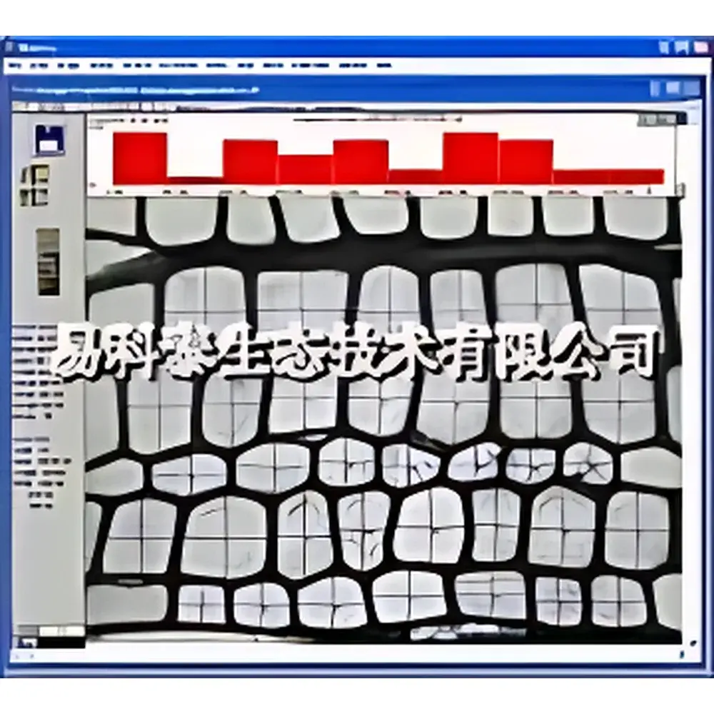
The WinCELL Microscopic Image Analysis System is a purpose-built, research-grade platform engineered specifically for quantitative dendroanatomical analysis of wood microstructure. Unlike generic image analysis tools, WinCELL integrates domain-specific algorithms rooted in tree-ring biology and xylem anatomy to deliver morphometric measurements that are statistically robust and biologically meaningful for dendrochronology, dendroclimatology, and wood science applications. The system operates on high-resolution optical microscopy images—acquired either from thin sections prepared with a microtome or directly from polished wood surfaces—and applies calibrated pixel-based segmentation to extract geometric and spatial parameters of tracheids (in conifers) and vessels (in angiosperms). Its core measurement principle relies on precise boundary detection of lumen contours and cell wall outlines, followed by topology-aware quantification that respects anatomical conventions—such as bisecting shared radial walls between adjacent cells when computing individual cell length or earlywood width. This ensures compliance with internationally recognized standards for anatomical wood measurement (e.g., IAWA Guidelines for Wood Anatomy, 2014; TRiDaS v2.0 metadata schema).
Key Features
- Domain-optimized analysis engine: Implements anatomically validated rules for cell wall partitioning, lumen classification, and radial row alignment—critical for accurate earlywood/latewood differentiation and functional position mapping along the growth ring.
- Multi-mode measurement architecture: Supports fully automated, semi-automatic (trace-guided), and interactive (point-and-click or line-drawing) analysis modes—enabling rigorous validation and correction where image contrast or section quality limits full automation.
- Flexible region-of-interest (ROI) definition: Standard version processes full-frame images; Enhanced version permits polygonal, rectangular, or circular ROI selection—essential for isolating specific growth rings, earlywood zones, or tangential bands for targeted analysis.
- Precision lumen and cell wall area quantification: Computes true pixel-based lumen area (not diameter-derived estimates) and wall area per cell, with optional classification into vessel vs. tracheid cohorts based on user-defined area thresholds.
- Color-coded lumen/wall area mapping (Enhanced version only): Enables spatial visualization of intra-ring variation in lumen area or wall thickness at the single-cell level—facilitating correlation with climate signals or mechanical property modeling.
- Cell position tracking relative to ring boundaries: Records Cartesian coordinates of cell centroids referenced to user-defined pith-to-bark axis, supporting statistical analysis of radial gradients in cell size or density across annual increments.
Sample Compatibility & Compliance
WinCELL accepts digitized images from brightfield or transmitted-light microscopy, including those acquired via standard C-mount adapters on compound or stereomicroscopes (Olympus, Leica, Zeiss, Nikon). Compatible sample formats include: (1) transverse microtome sections (5–20 µm thick), optionally stained with safranin-fast green or toluidine blue to enhance lumen-wall contrast; and (2) surface-imaged hardwood vessels (e.g., ring-porous species) under optimized oblique illumination. The system complies with FAO/IUFRO wood anatomy reporting conventions and supports metadata tagging aligned with TRiDaS v2.0 for interoperability with international dendro databases (e.g., ITRDB, TreeRing.org). All measurement workflows are traceable and reproducible—meeting GLP-aligned documentation requirements for environmental proxy studies.
Software & Data Management
WinCELL software (Standard or Enhanced edition) is TWAIN-compliant and interfaces natively with IEEE-1394 (FireWire) and USB 2.0 scientific cameras (1–6 MP resolution). Image acquisition is real-time or near-real-time, with adjustable exposure, gamma, and contrast settings prior to capture. Post-acquisition, raw TIFF or BMP files are processed without lossy compression. Measurement data—including lumen area, wall area, cell length/width, count per ROI, and centroid coordinates—are exported in tab-delimited ASCII format for downstream statistical analysis in R, Python (pandas), or MATLAB. Companion software XLCell provides advanced visualization: scatterplots of cell dimensions versus radial position, histograms of lumen area distributions per ring, and overlay heatmaps of spatial parameter variation. Audit trails, user session logs, and parameter configuration snapshots are retained for method validation and regulatory review (aligned with ISO/IEC 17025 documentation expectations).
Applications
- Dendroclimatology: Quantifying intra-annual xylem anatomy (e.g., vessel size, density, and arrangement) as high-resolution climate proxies—particularly sensitive to spring temperature and soil moisture availability.
- Wood quality assessment: Correlating tracheid/vessel morphology (lumen area, wall thickness, radial diameter) with mechanical properties (modulus of elasticity, compressive strength) and pulp yield potential.
- Tree physiology studies: Investigating cambial activity patterns, carbon allocation shifts, and stress responses (drought, frost, pollution) through temporal changes in cell differentiation dynamics.
- Forensic wood identification: Supporting species-level discrimination via statistically validated anatomical metrics (e.g., vessel grouping patterns, intervessel pit morphology).
- Educational use: Teaching plant anatomy, quantitative microscopy, and open-science data practices in undergraduate and graduate botany curricula.
FAQ
Does WinCELL require a specific microscope model?
No—WinCELL is microscope-agnostic and works with any compound or stereo microscope equipped with a C-mount port and appropriate camera adapter (sold separately by microscope manufacturers).
Can WinCELL analyze images from flatbed scanners?
Yes, but only for large-diameter vessels (e.g., oak, ash) where surface imaging yields sufficient contrast; scanner resolution must exceed 1200 dpi, and optical calibration is required.
Is FDA 21 CFR Part 11 compliance supported?
While WinCELL itself does not include electronic signature modules, its audit trail functionality (session logs, parameter history, export timestamps) forms a foundational layer for laboratories implementing Part 11–compliant SOPs in regulated environments.
What file formats does WinCELL export?
Measurement results are exported as plain-text tab-delimited (.txt) files compatible with Excel, R, and statistical packages; images are saved in lossless TIFF or BMP format.
Is training provided with system purchase?
Yes—Regent includes comprehensive documentation (printed manual + PDF), video tutorials, and up to two years of email-based technical support for method development and troubleshooting.


