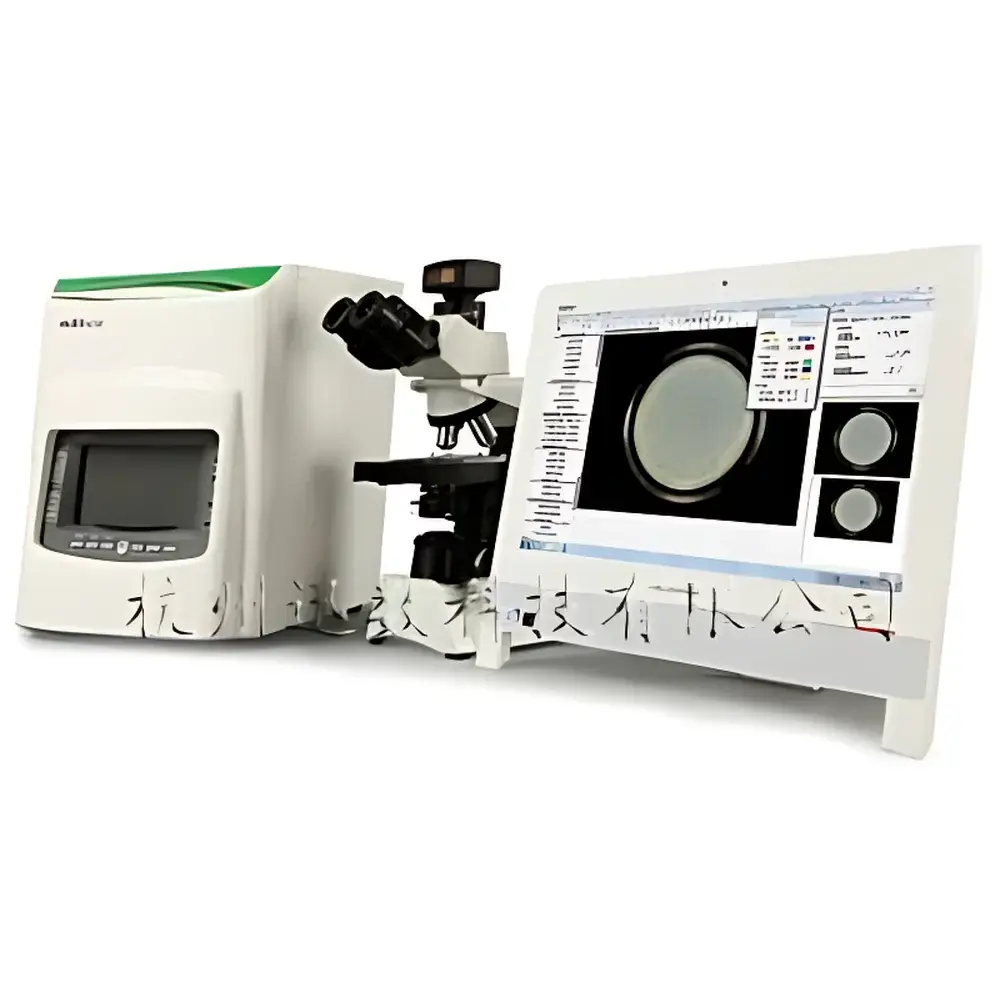
Xunsu MF1 Microbial Imaging & Analysis Workstation
| Brand | Xunsu |
|---|---|
| Origin | Zhejiang, China |
| Manufacturer Type | Direct Manufacturer |
| Instrument Type | Fully Automated Colony Counter |
| Counting Speed | <1 second for up to 500 colonies |
| Camera Resolution | 5 MP CMOS |
| Microscope Integration | Domestic upright microscope |
| UV Source | 254 nm LED |
| Illumination | Tri-color LED (white), adjustable top/bottom/dual lighting |
| Software Modules | Colony counting, Szone multi-mode zone-of-inhibition measurement, antibiotic potency calculation (ChP 2010), β-lactamase detection (Subactam-sensitive), and microscopic image analysis |
Overview
The Xunsu MF1 Microbial Imaging & Analysis Workstation is an integrated benchtop platform engineered for standardized, reproducible microbiological quantification and morphological analysis in academic teaching laboratories and QC environments. It combines digital colony imaging, high-fidelity抑菌圈 (zone-of-inhibition) metrology, and calibrated microscopic cell analysis within a single dark-box architecture. The system operates on the principle of high-contrast digital image acquisition under controlled illumination—leveraging tri-color visible-light LEDs and 254 nm UV for both disinfection and mutagenesis applications—and applies deterministic pixel-based segmentation algorithms to extract quantitative metrics from microbial growth patterns. Designed specifically for foundational life science education and routine pharmaceutical microbiology testing, the MF1 complies with core regulatory expectations for data integrity, traceability, and analytical transparency without requiring GMP-grade validation infrastructure.
Key Features
- Fully enclosed optical chamber with ergonomic viewing port eliminates ambient light interference, preventing halo artifacts caused by Petri dish refraction.
- Tri-color LED illumination system delivers accurate color rendition (CIE D65–compatible white balance), resolving spectral bias common in monochromatic white LEDs, while enabling selective contrast enhancement across bacterial pigments.
- Szone multi-mode zone-of-inhibition measurement engine implements three parallel edge-detection strategies: (1) contour-based automatic detection for crisp circular zones; (2) least-squares circular approximation for fragmented or irregular zones; and (3) manual three-point circle fitting for low-contrast or diffused boundaries.
- Integrated 5-megapixel industrial CMOS camera with fixed-focus lens ensures consistent spatial calibration across repeated acquisitions; supports real-time preview, frame capture, and lossless TIFF export.
- Dual-path lighting control: top-mounted 360° diffuse ring light for surface texture visualization; bottom-mounted “crystal-sharp” dark-field illumination for translucent colony differentiation on agar.
- Three-tier password protection (application launch, database access, administrator privileges) enforces role-based data governance aligned with GLP documentation practices.
- Onboard image processing suite includes 27 algorithmic tools—adaptive histogram equalization, morphological operators (erosion/dilation), Sobel/Canny edge detection, Gaussian/median filtering, and RGB channel-specific enhancement—optimized for biological specimen clarity.
Sample Compatibility & Compliance
The MF1 accommodates standard microbiological sample formats: pour plates, spread plates, and membrane filters (with grid-background suppression). Its colony counting module supports size-, shape-, and color-based classification across 25 diameter bins, with automated de-clumping for chain-forming organisms and user-guided separation for ambiguous clusters. For antibiotic susceptibility testing, it conforms to Chinese Pharmacopoeia (ChP) 2010 requirements for two-dose and three-dose potency assays—including merged calculation protocols—and provides built-in self-validation routines: repeatability error ≤0.01%, uniformity error ≤0.05%, inter-unit measurement deviation ≤0.2 mm. The β-lactamase detection module follows a validated Subactam-sensitive assay protocol using parallel disk diffusion controls (A/B/C/D), automatically computing mean zone diameters and issuing validity alerts when criteria (e.g., D−C ≥ 3 mm, B−A ≤ 3 mm) are unmet. All measurement metadata—including timestamps, operator IDs, instrument parameters, and raw image hashes—is embedded in exported Excel reports to support audit readiness.
Software & Data Management
The MF1 runs a unified software environment comprising five interoperable modules: ColonyCounter Pro, Szone Zone Analyzer, PotencyCalc (ChP-compliant), β-LactamaseAssay, and MicroScopeAnalyzer. Each module maintains independent audit trails compliant with FDA 21 CFR Part 11 principles: electronic signatures, immutable activity logs, and revision-controlled parameter histories. Statistical outputs—including total CFU, size distribution histograms, zone diameter matrices, and morphometric descriptors (circularity, aspect ratio, Feret diameter)—export directly to Microsoft Excel (.xlsx) with customizable field mapping. Image annotation tools permit overlay of scale bars, angular measurements, freehand curves, and typographic labels—facilitating direct integration into peer-reviewed manuscripts. Calibration is performed via on-screen reference targets or user-defined stage micrometer inputs, ensuring traceable dimensional accuracy down to 0.5 µm/pixel at default magnification.
Applications
The MF1 serves as a pedagogical and operational hub for undergraduate microbiology labs, food safety screening, dairy quality assurance, and early-stage pharmaceutical stability testing. Typical use cases include: enumeration of aerobic mesophiles in environmental swabs; quantitative assessment of antimicrobial efficacy against Staphylococcus aureus ATCC 25923; determination of penicillin G potency in bulk drug substances; verification of β-lactamase presence in raw milk samples; and morphometric profiling of yeast budding dynamics or filamentous fungal hyphae. Its modular design allows instructors to isolate specific workflows—e.g., restricting students to colony counting mode while reserving Szone and potency modules for advanced labs—without hardware reconfiguration.
FAQ
Does the MF1 meet regulatory requirements for pharmaceutical testing?
Yes—the system supports ChP 2010-compliant antibiotic potency calculations, includes built-in repeatability and uniformity self-tests, and generates audit-ready Excel reports with embedded metadata. While not pre-validated for GMP production environments, its architecture aligns with ALCOA+ data integrity principles.
Can the MF1 analyze Gram-stained or fluorescently labeled specimens?
It supports brightfield microscopy only; fluorescence or phase-contrast capability requires external microscope integration. However, its RGB-adjustable image enhancement tools improve contrast in stained preparations.
Is UV disinfection sufficient for complete sterilization of the imaging chamber?
The 254 nm UV source reduces surface bioburden effectively between runs but does not replace chemical decontamination for critical applications involving spore-forming organisms.
How is calibration maintained across long-term usage?
The system stores both factory-default and user-updated calibration coefficients. Operators may perform on-the-fly recalibration using certified stage micrometers or printed reference grids, with all changes logged in the audit trail.
What file formats does the software support for image import/export?
Native acquisition saves in lossless TIFF; exports include PNG, JPEG, BMP, and PDF (for annotated reports). Raw pixel data and measurement tables export exclusively to Excel (.xlsx) with column headers mapped to ISO/IEC 17025 reporting conventions.


